
Email id: info@ijclinmedcasereports.com
Intraoral Surgical Management of an Epidermoid Cyst of the Floor of the Mouth: A Case Report and Literature Review
Jabri M*, Hamza G, Lahjaouj M, Loudghiri M, Bijou W, Oukessou Y, Rouadi S, Abada RL, Roubal M and Mahtar M
ENT Department, 20th August 1953 Ibn Rochd University Hospital, Casablanca, Morocco
Received Date: 13/07/2026; Published Date: 21/10/2026
*Corresponding author: Jabri M, ENT Department, 20th August 1953 Ibn Rochd University Hospital, Casablanca, Morocco
Abstract
Background: Epidermoid cysts of the floor of the mouth are rare benign lesions arising from ectodermal entrapment during embryogenesis. Their anatomical relationship with surrounding muscles determines the surgical approach
Case Presentation: A 19-years-old male patient presented with a progressively enlarging swelling in the floor of the mouth. Cervicofacial CT scan revealed a well-defined cystic lesion above the mylohyoid muscle.
Complete surgical excision was performed via an intraoral approach. Histopathological examination confirmed an epidermoid cyst without malignancy.
The postoperative course was uneventful, with no recurrence on follow-up.
Conclusion: The intraoral approach is safe and effective for epidermoid cysts located above the mylohyoid muscle, providing excellent functional and cosmetic outcomes
Keywords: Epidermoid cyst; Floor of mouth; Intraoral approach; Dermoid cyst; Oral surgery
Introduction
Epidermoid cysts are benign developmental lesions resulting from ectodermal tissue entrapment during the fusion of the first and second branchial arches [1].
Histologically, they are lined by keratinized stratified squamous epithelium without skin appendages, differentiating them from dermoid and teratoid cysts [2].
Only 1.6–7% of epidermoid cysts occur in the head and neck region, and localization in the floor of the mouth is particularly rare [3,6].
Depending on their anatomical position, these lesions may involve the sublingual, submandibular, or submental spaces [4].
Clinically, they present as slow-growing, painless swellings but may lead to dysphagia, dysarthria, or airway compromise when enlarged [5,7]. Imaging modalities such as CT and MRI are essential for diagnosis and surgical planning [8].
Surgical excision is the treatment of choice. The approach depends on the relationship of the cyst to the mylohyoid and geniohyoid muscles [9]. We report a case managed successfully via an intraoral approach.
This case report has been reported in line with the SCARE checklist [1].
Case Report
A 19-years-old male presented with a midline swelling in the floor of the mouth evolving over four months. The patient reported mild discomfort during speech and swallowing. Clinical examination revealed a soft, non-tender mass elevating the tongue, with normal overlying mucosa (Figure 1).
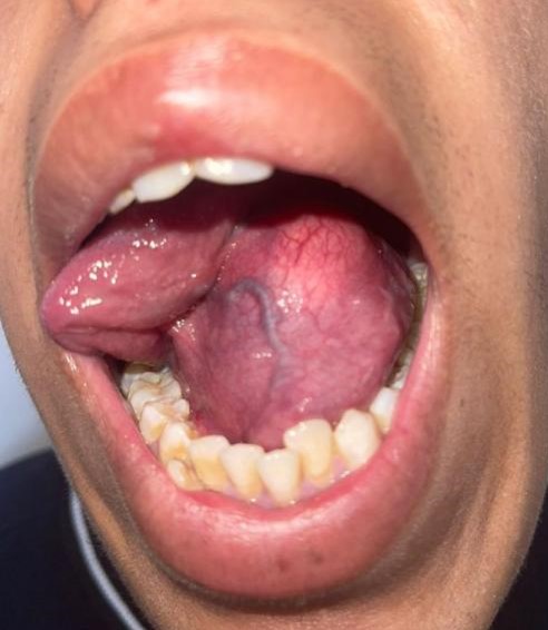
Figure 1: The clinical aspect of the mass of the mouth floor.
CT imaging demonstrated a well-circumscribed hypodense lesion located between the mylohyoid muscleand the geniohyoid muscle, suggestive of an epidermoid cyst (Figure 2).
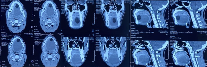
Figure 2: CT-Scan showing the the cystic formation of the mouth floor measuring 73,6 x 53,4 x 55,5 mm.
Given its supramylohyoid location, an intraoral surgical approach was selected (9,10). Under general anesthesia, a midline mucosal incision was made, and careful dissection allowed complete enucleation of the cyst while preserving adjacent structures (Figure 3).
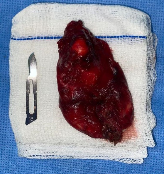
Figure 3: The completely excised epidermoid cyst.
Histopathological examination confirmed an epidermoid cyst characterized by keratinized stratified squamous epithelium and keratin content, without dysplasia or malignancy [2].
The postoperative course was uneventful. No complications or recurrence were observed during follow-up (Figure 4).
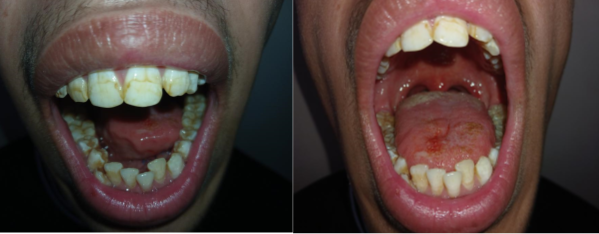
Figure 4: Post operative aspect of the mouth.
Discussion
Epidermoid cysts of the floor of the mouth are rare benign lesions that may present a diagnostic and therapeutic challenge due to their deep anatomical location and nonspecific clinical presentation [6].
They arise either from congenital ectodermal entrapment during embryologic fusion or, less commonly, from acquired causes such as traumatic or iatrogenic implantation of epithelial cells [2,6].
Clinically, these lesions typically manifest as slow-growing, painless swellings in the midline of the floor of the mouth.
However, depending on their size and location, they may lead to functional symptoms including dysphagia, dysarthria, tongue elevation, and, in severe cases, airway compromise [5,7].
In our case, the patient presented with mild functional discomfort without respiratory distress, which is consistent with early-stage lesions.
The differential diagnosis is broad and includes ranula, thyroglossal duct cyst, lymphangioma, cystic hygroma, and benign or malignant salivary gland tumors [7].
Distinguishing between these entities is crucial, as management strategies differ significantly. For instance, ranulas are typically associated with salivary gland extravasation and may require marsupialization or gland excision, whereas epidermoid cysts require complete surgical enucleation.
Radiological imaging plays a central role in both diagnosis and preoperative planning. CT scans typically reveal well-circumscribed, hypodense, non-enhancing lesions, while MRI offers superior soft tissue resolution and can better delineate the lesion’s relationship with adjacent muscles and neurovascular structures [8]. MRI may also help differentiate epidermoid cysts from dermoid cysts based on internal content characteristics. In our case, CT imaging was sufficient to establish the diagnosis and determine the surgical approach.
The anatomical classification proposed in the literature, based on the relationship of the cyst to the mylohyoid and geniohyoid muscles, is fundamental in guiding surgical management [9,11]. Lesions located above the mylohyoid muscle (sublingual space) are generally approached intraorally, whereas those located below the geniohyoid muscle (submental or submandibular spaces) often require an extraoral cervical approach.
The intraoral approach offers several advantages, including the absence of visible scarring, reduced surgical morbidity, shorter operative time, and faster postoperative recovery [10,12].
For these reasons, it should be preferred whenever anatomically feasible. Nevertheless, this approach requires meticulous surgical technique due to the proximity of critical structures such as Wharton’s ducts, the lingual nerve, and the sublingual glands. Inadequate dissection may result in complications such as ductal injury, sensory deficits, or incomplete excision.
Traditionally, large cysts were considered indications for an extraoral approach due to concerns regarding limited exposure and risk of rupture. However, recent studies have demonstrated that even relatively large cysts can be successfully managed intraorally with careful dissection and adequate surgical expertise [10,12,13]. This trend reflects a shift toward minimally invasive techniques in head and neck surgery.
Complete surgical excision remains the gold standard treatment, as partial removal or rupture of the cyst wall may increase the risk of recurrence. Fortunately, recurrence rates are low when complete enucleation is achieved [14]. Malignant transformation of epidermoid cysts is exceedingly rare but has been reported in the literature, emphasizing the importance of histopathological examination [14].
In our case, the lesion was located between the mylohyoid muscle and the geniohyoid muscle, allowing successful management via an intraoral approach with excellent functional and aesthetic outcomes. No postoperative complications or recurrence were observed, supporting the safety and efficacy of this technique.
Conclusion
Epidermoid cysts of the floor of the mouth should be considered in the differential diagnosis of midline oral swellings. Imaging is essential for diagnosis and surgical planning. The intraoral approach is a safe, effective, and cosmetically superior option for lesions located above the mylohyoid muscle, with excellent outcomes and low recurrence rates.
References
- Kerwan A, Al-Jabir A, Mathew G, Sohrabi C, Rashid R, Franchi T, et al. Revised Surgical CAse REport (SCARE) guideline: An update for the age of Artificial Intelligence. Premier Journal of Science, 2025; 10: 100079.
- Patil K, Mahima VG, Srikanth HS. Epidermoid cyst of floor of mouth: diagnostic challenges and management. J Oral Med Oral Surg, 2020; 26(1): 12-16.
- Rossi A, Di Nardo F, Boccuzzi A. Head and neck epidermoid cysts: clinical and histopathological features. Acta Otorhinolaryngol Ital, 2021; 41(2): 123-129.
- Boffano P, Roccia F, Gallesio C. Dermoid and epidermoid cysts of the oral cavity: clinical evaluation and management. J Craniofac Surg, 2021; 32(3): e234-e238.
- Shibata M, Ono S, Shinohara M. Surgical approaches to dermoid cysts of the floor of the mouth. Head Neck, 2021; 43(5): 1450-1456.
- Koca CF, Akcam T, Genc S. Sublingual epidermoid cysts: case series and literature review. Oral Maxillofac Surg, 2022; 26(2): 215-221.
- Kumar R, Singh AK, Mehrotra D. Management of dermoid cysts in the floor of mouth. Int J Oral Maxillofac Surg, 2022; 51(4): 512-518.
- Gupta A, Singh S, Kumar P. Rare presentations of epidermoid cysts in oral cavity. Case Rep Dent, 2021; 2021: 6632451.
- Uehara M, Takahashi Y, Satoh K. Imaging features of dermoid and epidermoid cysts in oral cavity. Radiol Case Rep, 2023; 18(1): 210-215.
- Yamamoto K, Kurihara K, Minami M. Clinical evaluation of dermoid cysts of the floor of the mouth. Med Oral Patol Oral Cir Bucal, 2020; 25(3): e391-e397.
- Singh V, Bhagol A, Kumar I. Intraoral excision of large epidermoid cysts: a safe approach. J Maxillofac Oral Surg, 2021; 20(2): 298-303.
- Chen S, Li Y, Wang X. Surgical management of dermoid cysts of the floor of the mouth. Oral Surg Oral Med Oral Pathol Oral Radiol, 2022; 133(4): e180-e186.
- Lee DH, Yoon TM, Lee JK. Minimally invasive intraoral approach for sublingual cysts. Clin Exp Otorhinolaryngol, 2022; 15(2): 200-205.
- Zhang W, Chen Y, Liu Y. Surgical outcomes of floor of mouth cysts: a retrospective study. BMC Oral Health, 2023; 23(1): 145.
- Al-Khateeb TH, Al-Masri NM. Dermoid cysts of the floor of the mouth: long-term outcomes and recurrence. J Oral Maxillofac Surg, 2020; 78(9): 1545-1552.