
Email id: info@ijclinmedcasereports.com
Spontaneous Remission of Type 2 Autoimmune Pancreatitis: A Case of Complete Imaging Reversal
Leandros Sassis, Pelagia Kefala Karli, Elena Xenofontos, Loukia Dramiotou and Maria Skarpari*
Department of Internal Medicine, Limassol General Hospital, State Health Services Organization, Limassol, Cyprus
Received Date: 23/07/2026; Published Date: 25/09/2026
*Corresponding author: Maria Skarpari, Department of Internal Medicine, Limassol General Hospital, State Health Services Organization, Limassol, Cyprus
Abstract
Background: Autoimmune Pancreatitis (AIP) is defined as a chronic autoimmune disease-causing inflammation of the pancreas. Nowadays, the diagnostic evaluation of AIP type 2 remains difficult due to a lack of specific serological and imaging findings in cases where biopsy is inconclusive. Corticosteroids are the mainstay of treatment for autoimmune pancreatitis; however, their use should be individualized, as treatment is primarily indicated in symptomatic patients, while selected asymptomatic cases may be managed conservatively.
Case report: We report a 34-year-old patient who presented with sudden abdominal pain and vomiting with no precipitating factors. Computed Tomography (CT) revealed typical findings of acute pancreatitis. Despite optimal management based on the provisional diagnosis, the patient developed obstructive jaundice, fever, and increased inflammation markers. MRI abdomen imaging revealed a “sausage-shaped” pancreas, a sign compatible with AIP. IgG4 antibodies were negative, and follow-up abdominal MRI showed complete resolution of prior findings without immunosuppressive therapy. The presence of coexisting ulcerative colitis serves as an important supportive feature for the diagnosis of type 2 AIP.
Conclusion: Although corticosteroids represent the standard therapy choice, type 2 AIP may follow a self-limited course, as illustrated in our case, with complete clinical and radiological resolution in the absence of treatment. This highlights the importance of careful patient selection and consideration of a conservative approach in selected cases to avoid unnecessary treatment or surgical intervention. Further studies are needed to identify predictors of spontaneous remission and to clarify the role of diagnostic modalities in guiding early and accurate diagnosis.
Keywords: Autoimmune pancreatitis type 2; Spontaneous remission; Sausage-shaped pancreas; Ulcerative colitis
Introduction
Autoimmune Pancreatitis (AIP) is a rare form of chronic pancreatic inflammation caused by immune response dysfunction, where the patient’s immune system targets healthy pancreatic tissue and is usually well responsive to steroid therapy. Nowadays, AIP is classified into three subtypes with distinct clinical and pathological features; Type I AIP (IgG4-related pancreatitis), type II AIP (Idiopathic duct-centric pancreatitis), and type III AIP (Immune checkpoint inhibitor–induced AIP) [1].
Type II AIP is confined to the pancreatic tissue, and there is no involvement of other organs, nor elevated serum IgG4 levels compared to type I. Several other antibodies have been proposed as potential markers for AIP type 2, including serum myeloperoxidase antibodies, anti-smooth muscle antibodies, anti-trans aldolase antibodies, anti-amylase antibodies, and perinuclear or cytoplasmic antineutrophil cytoplasmic antibodies. The latter are also commonly found in patients with ulcerative colitis; however, the diagnostic value of these markers require further validation [2].
Typical clinical features are abdominal pain with a lower proportion of the presence of jaundice and recurrent episodes of acute pancreatitis with low-grade severity [2]. It usually affects middle-aged men and women in equal proportion, with almost half of the patients suffering with inflammatory bowel disease, too [1]. Histological findings are essential for the diagnosis of AIP type 2. In cases where histology is inconclusive or not performed, the presence of ulcerative colitis, supports a diagnosis of “probable type 2 AIP” [3].
Although corticosteroids are considered the standard treatment, the natural history of type 2 AIP is not fully understood. In the literature, few cases of spontaneous remission have been described but remains uncommon, and predictors of such outcomes are lacking [4]. We herein report a case of type 2 AIP presenting with obstructive jaundice and spontaneous resolution, underscoring the ongoing diagnostic challenges and the need for a more individualized therapeutic approach.
Case Report
A 34-year-old Indian male with no significant medical history presented to the ER complaining of persistent epigastric abdominal pain, not associated with food, and refused alcohol intake, radiating to the back that started 24 hours before admission, and one episode of non-bloody vomit. The physical examination revealed abdominal sensitivity/tenderness in the epigastric and peri-omphalic area, along with guarding on deep palpation; bowel sounds were normal. From the rest of the physical examination, there were no other abnormal findings, and the vital signs were normal.
Laboratory findings on admission revealed increased inflammation markers, serum amylase (2,036 reference value: 28-100 U/L) and urine amylase (19,451 reference value: 21-447 U/L) as well as liver and cholestatic enzymes. Laboratory values and trends of the liver-cholestatic enzymes are displayed on Figure 1.
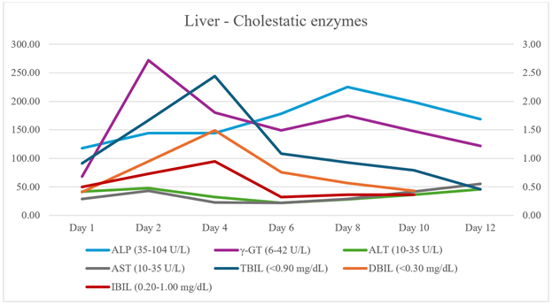
Figure 1: Laboratory values and trends of the liver-cholestatic enzymes during patient’s hospitalization.
Computed Tomography (CT) of the abdomen and pelvis with IV contrast (Figure 2A) was performed and showed significant pancreatic edema and peri-pancreatic fluid collection, diffuse enlargement of the pancreatic gland, and blurring of peripancreatic fat, whereas chest x-ray did not show any abnormal findings.
The patient was admitted to the internal medicine ward, and rigorous hydration was administered. On further laboratory investigation, hypertriglyceridemia and hypercalcemia were excluded. Further investigation was performed, including viral blood test (CMV, HAV, HBV, HBC, HIV, Rubella) and test for Toxoplasma gondii, which were negative, and autoimmune blood tests for IgG4 levels were performed, although the results were not available during his hospitalization.
On the fourth day of hospitalization, the patient got febrile, and he had several episodes of watery diarrhea; on clinical examination, tenderness in the right upper quadrant with Murphy’s sign was elicited, and the laboratory tests revealed increased inflammation markers and cholestatic enzymes (Figure 1). Blood, urine, and stool cultures were obtained which were negative. An emergency liver/bile ducts ultrasound was performed which showed gallbladder wall thickening without evidence of cholelithiasis or bile duct distention, with the presence of a small collection of peripancreatic fluid. Thus, Magnetic Resonance Imaging (MRI) of the abdomen with IV contrast confirmed acute inflammation of the pancreatic tissue. The report highlighted the gross enlargement of pancreatic parenchyma with restricted diffusion and homogenous filling described as “sausage”-shaped pancreas, with diffused stenosis of the pancreatic duct and peripancreatic oedema and without obvious presence of pancreatic pseudocysts (Figure 2Β).
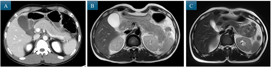
Figure 2: A) Contrast-enhanced CT of the abdomen and pelvis showing diffuse pancreatic enlargement with edema, peripancreatic fluid, and fat blurring, B) Abdominal MRI (T2) showed “sausage-shaped” pancreas; enlargement of pancreatic parenchyma with restricted diffusion and homogenous filling, C) Follow-up abdominal MRI (T2) showed complete resolution of the findings of the initial MRI.
In the context of clinical and laboratory features suggestive of acute cholecystitis, empirical treatment with imipenem/cilastatin was initiated. Following clinical improvement of epigastric pain, oral feeding started with no abdominal discomfort. During hospitalization, the patient showed clinical and laboratory improvement, and then he left against medical advice before the results of IgG4 levels were available. Only dietary recommendations were given, and a follow-up appointment was arranged.
On outpatient follow-up, immunological testing for the presence of IgG4 antibodies was negative, and a repeat abdominal MRI performed three months after the initial episode demonstrated complete resolution of the previously observed findings (Figure 2C). Furthermore, colonoscopy revealed features consistent with ulcerative colitis. Thus, the diagnosis of type 2 autoimmune pancreatitis (AIP) with spontaneous remission was suspected based on the combination of clinical presentation, imaging resolution, and the associated diagnosis of ulcerative colitis.
Discussion
Due to its rarity, little is known about AIP type 2, in contrast to the substantially better-established understanding of AIP type 1, which has evolved considerably over time. Diagnostic criteria of AIP are based on a combination of clinical presentation, serological markers, imaging findings, histology, and response to corticosteroids [3]. Despite these criteria, the diagnosis of AIP-2 remains challenging, largely due to its low prevalence, limited histological sampling, and absence of reliable serum biomarkers.
Except for indistinguishable imaging signs and a well-established respond on steroids, AIP type 2 generally shows different clinical, histologic, and prognostic characteristics from AIP type 1[1]. Clinically, type 2 AIP differs from type 1 in that serum IgG4 levels are typically normal or low, and there is usually no other-organ involvement [1]. However, inflammatory bowel disease (IBD), particularly ulcerative colitis, is associated in approximately 15–45% of cases [5]. This association represents one of the few consistent extra-pancreatic features of type 2 AIP.
Histopathology remains the gold standard for diagnosis. It is characterized by a duct-centric fibro-inflammatory process with neutrophilic infiltration of medium and small pancreatic ducts and the presence of granulocytic epithelial lesions, which represent its most specific histopathological feature [1,5]. However, pancreatic biopsy is not always feasible in clinical practice due to its invasive nature and associated procedural risks. This limitation is particularly relevant in suspected AIP-2, where tissue sampling is often unavailable, further complicating definitive diagnosis.
The absence of reliable serological markers and the limited availability of histological confirmation frequently result in delayed diagnosis, misclassification, or unnecessary surgical intervention. In clinical practice, a probable diagnosis of AIP-2 is often made by integrating imaging findings, steroid responsiveness, and the presence of concomitant IBD when histology is unavailable or non-diagnostic. This pragmatic approach is supported by international consensus recommendations [3].
Radiologically, as seen in our case, focal AIP may demonstrate a range of characteristic findings, including sausage-shaped enlargement, delayed homogeneous enhancement, a capsule-like rim, irregular narrowing of the main pancreatic duct (MPD) and/or common bile duct strictures, and upstream MPD dilation ≤5 mm [6]. Importantly, sausage-shaped enlargement was observed in 11 of 19 focal AIP cases but was not seen in pancreatic cancer, supporting its diagnostic specificity [6]. This imaging feature has been consistently reaffirmed in subsequent studies as a key characteristic of AIP [7].
Corticosteroids remain the cornerstone of therapy for autoimmune pancreatitis. Standard treatment typically consists of oral prednisone at a dose of 40 mg daily for 4–6 weeks, followed by a gradual taper of approximately 5 mg per week over a two-month period, although the optimal duration of therapy and long-term outcomes remain uncertain [8]. In patients with relapse or steroid dependence, rituximab may be considered as an alternative therapeutic option, while steroid-sparing agents such as thiopurines have demonstrated limited efficacy [9].
According to the 2017 International Consensus, indications for steroid therapy include symptomatic pancreatic disease such as obstructive jaundice and abdominal pain, involvement of other organs, and persistent pancreatic masses or systemic manifestations consistent with IgG4-related disease [9]. Treatment is also indicated in patients with progressive disease affecting critical structures, including biliary obstruction, cholangitis, or evidence of irreversible organ damage such as pancreatic exocrine or endocrine insufficiency and hepatobiliary fibrosis. A Japanese study from a long-term follow-up study of 23 patients with AIP concludes that the indications for steroid therapy in AIP are obstructive jaundice due to stenosis of the bile duct or other associated systemic autoimmune diseases, where the presence of diabetes mellitus might be an indication [8].
Despite the general effectiveness of corticosteroids, spontaneous remission has been reported in a subset of patients with AIP, particularly in type 2 disease. AIP-2 is treated less frequently with corticosteroids compared with type 1 disease and demonstrates lower relapse rates (9% and 31%, respectively) [9]. Moreover, remission rates appear similar between treated and untreated patients, suggesting that maintenance therapy may not be necessary in this subgroup [9].
Several studies support the phenomenon of spontaneous remission. Kamisawa et al. [8] reported spontaneous improvement in 3 of 23 patients, while 10 underwent surgical intervention. Notably, 50% of patients treated with corticosteroids developed pancreatic atrophy, highlighting potential treatment-related adverse effects and raising questions about overtreatment in selected cases [8]. Similarly, Kubota et al. [10] reported spontaneous remission in 65% of untreated patients and identified low disease activity and IgG4 seronegativity as independent predictors of spontaneous remission. Additional favorable prognostic factors included the absence of obstructive jaundice or of diabetes mellitus, the lack of duodenal papillary swelling, negative IgG4 staining of the duodenal papilla, and focal pancreatic involvement on imaging. Among additional histological and endoscopic markers, negative IgG4 staining of the duodenal papilla has been proposed as a potential predictor of spontaneous remission. However, its independent predictive value remains limited [10].
Based on current evidence, a conservative “watch-and-wait” approach may be appropriate in selected asymptomatic patients, particularly those without papillary swelling and with favorable imaging features. This strategy is supported by International Consensus guidelines which recommend observational management in carefully selected cases [9].
In the present case, both clinical symptoms and imaging abnormalities resolved completely without pharmacological intervention, as the patient left against medical advice. This spontaneous resolution further supports the recognized benign and reversible nature of type 2 AIP. In contrast to type 1 disease, cross-sectional imaging in type 2 AIP -particularly MRI performed after the acute inflammatory phase- may appear normal, as the inflammatory process is typically duct-centric and does not usually result in significant fibro-inflammatory remodeling. Consequently, structural abnormalities may resolve entirely without residual fibrosis on follow-up imaging. This reflects the generally benign and reversible nature of type 2 AIP and supports the observation that imaging can return to baseline in the absence of chronic fibrotic remodeling [7].
Conclusion
Although diagnostic criteria are well-established, AIP type 2, due to its rarity, is not easily diagnosed, especially in cases where biopsy is not feasible to perform. Evaluation of clinical, imaging, and colonoscopy for the diagnosis of IBD is crucial for the diagnostic approach. Moreover, the role of corticosteroid therapy remains challenging, given the occurrence of spontaneous remission in some patients and the well-recognized risk of treatment-related complications. Improvement of the current imaging modalities and novel biomarkers need to be developed to enhance early and accurate diagnosis of AIP type 2 and prevent unnecessary diagnostic and therapeutic interventions.
Learning points:
- Type 2 autoimmune pancreatitis can present without typical serological markers, making diagnosis reliant on clinical context and imaging findings.
- Spontaneous remission of Type 2 autoimmune pancreatitis may occur without corticosteroid therapy.
- Association with inflammatory bowel disease, particularly ulcerative colitis, can provide an important diagnostic clue in suspected Type 2 autoimmune pancreatitis.
Disclosure
Conflict of interest: The authors declare no conflict of interest.
Patient Consent: Written informed consent was obtained from the patient for publication of this case report and accompanying clinical details.
Acknowledgments: The authors acknowledge the medical and nursing staff involved in the patient’s care for their valuable support and contributions.
References
- Vemulapalli V, Natha C, Shirwaikar Thomas A, Autoimmune Pancreatitis: A Review. J Clin Med, 2025; 14. doi: 10.3390/jcm14093076.
- Li Y, Song H, Meng X, Li R, Leung PSC, Gershwin ME, et al. Autoimmune pancreatitis type 2 (idiopathic duct-centric pancreatitis): A comprehensive review. J Autoimmun, 2023; 140: 103121. doi: 10.1016/j.jaut.2023.103121.
- Shimosegawa T, Chari ST, Frulloni L, Kamisawa T, Kawa S, Mino-Kenudson M, et al. International consensus diagnostic criteria for autoimmune pancreatitis: guidelines of the International Association of Pancreatology. Pancreas, 2011; 40: 352-358. doi: 10.1097/MPA.0b013e3182142fd2.
- Zhang BB, Huo JW, Yang ZH, Wang ZC, Jin EH, Spontaneous remission of autoimmune pancreatitis: Four case reports. World J Clin Cases, 2022; 10: 8232-8241. doi: 10.12998/wjcc.v10.i23.8232.
- Lee TH. Differentiation of Type 2 Autoimmune Pancreatitis Presenting as Clinical Acute Pancreatitis. Gut Liver, 2019; 13: 385-387. doi: 10.5009/gnl19175.
- Sun GF, Zuo CJ, Shao CW, Wang JH, Zhang J. Focal autoimmune pancreatitis: radiological characteristics help to distinguish from pancreatic cancer. World J Gastroenterol, 2013; 19: 3634-3641. doi: 10.3748/wjg.v19.i23.3634.
- Slaoui H, Beyyato S, Chaouche I, Ouazzani H, Marghich O, Akammar A, et al. Autoimmune pancreatitis: A series of 10 cases. Medical Reports, 2025; 13: 100324. doi: https://doi.org/10.1016/j.hmedic.2025.100324.
- Kamisawa T, Yoshiike M, Egawa N, Nakajima H, Tsuruta K, Okamoto A. Treating patients with autoimmune pancreatitis: results from a long-term follow-up study. Pancreatology, 2005; 5: 234-240. doi: 10.1159/000085277.
- Okazaki K, Chari ST, Frulloni L, Lerch MM, Kamisawa T, Kawa S, et al. International consensus for the treatment of autoimmune pancreatitis. Pancreatology, 2017; 17: 1-6. doi: https://doi.org/10.1016/j.pan.2016.12.003.
- Kubota K, Iida H, Fujisawa T, Yoneda M, Inamori M, Abe Y, et al. Clinical factors predictive of spontaneous remission or relapse in cases of autoimmune pancreatitis. Gastrointestinal Endoscopy, 2007; 66: 1142-1151. doi: https://doi.org/10.1016/j.gie.2007.06.059.