
Email id: info@ijclinmedcasereports.com
Vancomycin-Induced Drug Reaction with Eosinophilia and Systemic Symptoms (DRESS) in a Patient with Infective Endocarditis: A Case Report and Contemporary Review of the Literature
Nikhilesh Anand1, Navin Patil2, Samith Ahmed3, Shanti Gurung4, Samira Abdul Wajid5,*, Pradeep Kumar Nambi6, Afreen Begum Hasansab Itagi7, Suraiya Abdul Wajid8, Parmida Sanaei9, Justin Singh9, Sudheeksha Banglore Venkatesh9 and Patricio Gratuito Jr10
1Department of Medical Education, University of Texas Rio Grande Valley, School of Medicine, Edinburg, Texas, USA
2Professor/Chair Department of Preclinical Sciences, Windsor University School of Medicine, St Kitts and Nevis
3Professor /Chair Department of Pathophysiology, Windsor University School of Medicine, St Kitts and Nevis
4Associate Professor, Department of Preclinical Sciences, Windsor University School of Medicine, St Kitts and Nevis
5Professor Microbiology and Director of SASP, Windsor University School of Medicine, St Kitts and Nevis
6Assistant Professor, Department of Anatomical Sciences, Windsor University school of Medicine, St Kitts and Nevis
7CPAC Clinician- Sleep Technicologist, Sleep disorders clinic, Hamilton-ON
8Postgraduate student Pathology, Srinivas Institute of Medical Sciences and Research Centre, Mangalore
9 Undergraduate student, Windsor University School of Medicine, St Kitts and Nevis
10Undergraduate MD5 student
Received Date: 03/07/2026; Published Date: 11/09/2026
*Corresponding author: Dr. Samira Abdul Wajid, MD, Professor of Microbiology and Director, Student Academic Support Program, Windsor University School of Medicine, Brighton's Estate, Cayon, St. Kitts and Nevis
Abstract
Background: Drug Reaction with Eosinophilia and Systemic Symptoms (DRESS), also termed Drug-Induced Hypersensitivity Syndrome (DIHS), is a severe, T-cell-mediated, delayed-type idiosyncratic adverse drug reaction characterised by a prolonged latency period (typically 2–8 weeks), morbilliform exanthem, fever, lymphadenopathy, haematologic abnormalities (eosinophilia and/or atypical lymphocytosis), and visceral organ involvement. Reported incidence ranges from 1 in 1,000 to 1 in 10,000 drug exposures, with case-fatality rates between 2 % and 10 %. Although classically associated with aromatic anticonvulsants, allopurinol and sulphonamides, vancomycin has emerged over the past decade as a leading antibiotic trigger, with the HLA-A*32:01 allele now established as a major genetic risk factor.
Case Presentation: A 61-year-old woman with type 2 diabetes mellitus and bronchial asthma was admitted with a one-week history of dyspnoea, cough and intermittent high-grade fever. Transthoracic and transoesophageal echocardiography demonstrated mobile vegetation on the ventricular surface of the anterior mitral leaflet, and a clinical diagnosis of culture-negative infective endocarditis was made. She received intravenous ceftriaxone (2 g once daily) and vancomycin (1 g twice daily) for an intended four-week course. On approximately the 24th day of vancomycin therapy she developed a generalised pruritic morbilliform eruption with facial oedema, high-grade fever (38.6 °C with chills and rigors), and laboratory evidence of peripheral eosinophilia, leukocytosis, lymphadenopathy and acute kidney injury. Concomitant medications were reviewed, alternative infectious and autoimmune aetiologies were excluded, and a punch skin biopsy demonstrated epidermal spongiosis with lymphocytic exocytosis and a dermal lymphocytic infiltrate containing eosinophils, consistent with a drug-induced spongiotic dermatitis. The RegiSCAR diagnostic criteria were fulfilled at the level of a probable case of DRESS.
Management and Outcome: Vancomycin was discontinued immediately; ceftriaxone was continued to complete antimicrobial coverage. First-generation H₁-antihistamines (pheniramine and hydroxyzine) produced no meaningful improvement, prompting initiation of systemic corticosteroid therapy. Cutaneous and constitutional symptoms resolved progressively, renal function normalised, and the patient was discharged in stable condition with structured outpatient follow-up. A causality assessment using the Naranjo Adverse Drug Reaction Probability Scale yielded a score in the “probable” range; the reaction was classified as severe (Hartwig Level 5) and not preventable (Schumock and Thornton criteria).
Conclusions: Vancomycin-induced DRESS is increasingly recognised in parallel with the global expansion of vancomycin use against methicillin-resistant Staphylococcus aureus and other Gram-positive infections, yet remains under-reported and frequently mis-diagnosed as red-man syndrome, sepsis, or other severe cutaneous adverse reactions. Clinicians prescribing prolonged vancomycin courses—particularly for endocarditis, osteomyelitis and bacteraemia—should maintain a high index of suspicion when fever, eosinophilia, transaminitis or acute kidney injury develop between two and eight weeks of therapy. Early withdrawal of the culprit drug and judicious systemic corticosteroid therapy remain the cornerstones of management.
Keywords: Vancomycin; DRESS syndrome; Drug-Induced Hypersensitivity Syndrome; Adverse cutaneous drug reaction; Infective endocarditis; HLA-A*32:01; RegiSCAR; Pharmacovigilance
Abbreviations: ACDR - Adverse Cutaneous Drug Reaction; ADR - Adverse Drug Reaction; AGEP - Acute Generalised Exanthematous Pustulosis; AKI - Acute Kidney Injury; AML - Anterior Mitral Leaflet; CARE - CAse REport (reporting guideline); CMV - Cytomegalovirus; DIHS - Drug-Induced Hypersensitivity Syndrome; DRESS - Drug Reaction with Eosinophilia and Systemic Symptoms; EBV - Epstein–Barr Virus; HHV-6 / HHV-7 - Human Herpesvirus 6 / 7; HLA - Human Leukocyte Antigen; IE - Infective Endocarditis; MRSA - Methicillin-resistant Staphylococcus aureus; NSAID - Non-Steroidal Anti-Inflammatory Drug; RegiSCAR - European Registry of Severe Cutaneous Adverse Reactions; SCAR - Severe Cutaneous Adverse Reaction; SJS - Stevens–Johnson Syndrome; TEN - Toxic Epidermal Necrolysis; TOE - Transoesophageal Echocardiography
Introduction
Adverse Drug Reactions (ADRs) are a leading iatrogenic cause of hospital admission and in-hospital morbidity worldwide, accounting for an estimated 3–6 % of all hospitalisations and contributing to approximately 100,000 deaths annually in the United States alone [1,2]. The skin is the organ most frequently affected, with cutaneous ADRs reported in 2–5 % of hospitalised patients in developed countries and in 2–6 % of outpatients in published Indian series [3]. Although the great majority of cutaneous reactions are benign and self-limiting, a small subset—collectively termed Severe Cutaneous Adverse Reactions (SCARs)—carry substantial morbidity and may be life-threatening. SCARs encompass Stevens–Johnson Syndrome (SJS), Toxic Epidermal Necrolysis (TEN), Acute Generalised Exanthematous Pustulosis (AGEP) and Drug Reaction with Eosinophilia and Systemic Symptoms (DRESS).
DRESS, first formally delineated by Bocquet and colleagues in 1996 and subsequently characterised in detail by the European Registry of Severe Cutaneous Adverse Reactions (RegiSCAR) consortium, is a severe T-cell-mediated, delayed-type idiosyncratic reaction with an incidence of approximately 1 in 1,000 to 1 in 10,000 drug exposures and a reported case-fatality rate of 2–10 % [4,5]. Its hallmark features include a long latency period (typically 2–8 weeks after drug initiation), an extensive morbilliform exanthem with facial oedema, high-grade fever, generalised lymphadenopathy, peripheral eosinophilia and/or atypical lymphocytosis, and involvement of one or more internal organs—most commonly the liver, but also the kidneys, lungs, heart, pancreas and central nervous system [6,7]. Long-term sequelae, including autoimmune thyroiditis, fulminant type 1 diabetes mellitus and myocarditis, may develop months after apparent clinical resolution [8].
The pathogenesis of DRESS is multifactorial. Three mutually compatible mechanisms are currently invoked: (i) accumulation of reactive drug metabolites in genetically susceptible individuals with impaired detoxification capacity; (ii) class I Human Leukocyte Antigen (HLA)–restricted, drug-specific CD8⁺ cytotoxic T-lymphocyte activation, often via the pharmacological interaction with immune receptors (“p-i”) concept proposed by Pichler; and (iii) reactivation of latent human herpesviruses—principally Human Herpesvirus 6 (HHV-6), but also HHV-7, Epstein–Barr virus and cytomegalovirus—which both perpetuates and amplifies the immune response [7,9,10]. Pharmacogenomic associations have been demonstrated for several culprit drugs, including HLA-B*15:02 and HLA-B*15:11 with carbamazepine, HLA-B*58:01 with allopurinol, and—of particular relevance to the present case—HLA-A*32:01 with vancomycin, which Konvinse and colleagues identified in 2019 with an estimated odds ratio of approximately 75 in patients of European ancestry [11].
Vancomycin, a tricyclic glycopeptide antibiotic introduced into clinical practice in 1958, remains a first-line agent for serious infections caused by Methicillin-Resistant Staphylococcus Aureus (MRSA), ampicillin-resistant enterococci, and selected Gram-positive organisms in patients with severe β-lactam allergy. Its consumption has increased markedly in the past two decades, in parallel with the global rise of MRSA. Although infusion-related histamine release (“red-man syndrome”), nephrotoxicity, ototoxicity and neutropenia are well-recognised adverse effects, vancomycin is now also acknowledged as one of the most frequently implicated antibiotics in DRESS, accounting for the majority of antimicrobial-attributable cases in several recent series [12–14]. Despite this, awareness among non-dermatology prescribers remains limited, and the literature is dominated by small case series and individual reports.
We present a clinically and pathologically substantiated case of vancomycin-induced DRESS in an elderly Indian woman treated for culture-negative infective endocarditis, and we contextualise the case with a contemporary, structured review of the published literature, including a comparative tabulation of representative reports. Reporting follows the CARE (CAse REport) guidelines [15].
Case Presentation
Presenting complaint and history:
A 61-year-old woman of South Indian ethnicity presented to the emergency department of a tertiary-care teaching hospital in southern India with progressive exertional dyspnoea of seven days' duration, accompanied by a productive cough and high-grade intermittent fever for three days. The fever was without diurnal variation, was associated with sweating and chills, and was transiently relieved by over-the-counter antipyretics. The patient denied chest pain, palpitations, orthopnoea, paroxysmal nocturnal dyspnoea, haemoptysis, peripheral oedema, weight loss, recent dental procedures, intravenous drug use or known cardiac disease.
Her past medical history was notable for type 2 diabetes mellitus, diagnosed approximately one year prior to admission and managed with oral hypoglycaemic therapy, and for bronchial asthma, diagnosed three months prior and treated with an inhaled short-acting β₂-agonist on an as-required basis. She denied previous adverse drug reactions, including to penicillins, cephalosporins or sulphonamides. She had no known food allergies, was a lifelong non-smoker, did not consume alcohol, and reported no recent travel, animal exposures or known sick contacts. Family and surgical histories were non-contributory.
Examination on admission:
On admission the patient was conscious, alert, and oriented to time, place and person. She was febrile but haemodynamically stable, with a blood pressure of 120/80 mmHg, a pulse rate of 80 beats per minute that was regular in rhythm, and oxygen saturation maintained on room air. There was no pallor, icterus, cyanosis, clubbing, peripheral lymphadenopathy or pedal oedema at the time of admission. Examination of the respiratory system revealed bilateral expiratory rhonchi, consistent with her known asthma. Cardiovascular examination did not initially disclose a clinically appreciable murmur. The abdomen was soft and non-tender, without organomegaly. Neurological examination was unremarkable, and there were no peripheral stigmata of infective endocarditis (no splinter haemorrhages, Janeway lesions, Osler nodes, or Roth spots).
Initial investigations and diagnosis of infective endocarditis:
Three sets of blood cultures, drawn from three separate venepuncture sites with an interval of at least one hour, returned no growth after standard incubation, consistent with culture-negative endocarditis—a recognised entity accounting for between 2.5 % and 31 % of cases of infective endocarditis in published series, particularly when antibiotics have been administered prior to culture or when fastidious or non-bacterial organisms are involved. Routine biochemistry, complete blood count, urinalysis, inflammatory markers, electrocardiography and chest radiography were performed at the time of admission as part of standard pre-treatment workup.
Transthoracic echocardiography demonstrated an oscillating, echogenic mass measuring approximately 3 × 2 mm attached to the atrial aspect of the base of the anterior mitral leaflet (AML), raising suspicion of vegetation. Subsequent transoesophageal echocardiography (TOE), which has superior sensitivity for native-valve vegetations, confirmed a mobile vegetation measuring approximately 2 × 1 mm on the ventricular surface of the AML, without significant mitral regurgitation, leaflet perforation or perivalvular abscess. According to the modified Duke criteria, the patient fulfilled one major criterion (echocardiographic evidence of endocardial involvement) and supportive minor criteria (predisposing factor in the form of diabetes mellitus, fever > 38 °C), and a diagnosis of possible-to-definite culture-negative infective endocarditis was made.
Antimicrobial therapy and clinical course:
Empirical antimicrobial therapy was initiated in accordance with contemporary guidelines for culture-negative native-valve endocarditis: intravenous ceftriaxone 2 g once daily and intravenous vancomycin 1 g twice daily (administered over at least 60 minutes each, with vancomycin serum trough monitoring as per institutional protocol). Inhaled bronchodilators and oral hypoglycaemic therapy were continued; no other new systemic medications were introduced during the first three weeks of admission. After two weeks of therapy, repeat blood cultures remained sterile and follow-up echocardiography demonstrated reduction in the size of the vegetation; in view of the diagnosis of infective endocarditis, the combination regimen was extended for an additional two weeks, for a planned total duration of four weeks.
Approximately ten days into the extended course—corresponding to the 24th day of vancomycin therapy overall—the patient developed a new pruritic erythematous eruption that began on the trunk and rapidly became generalised, involving the face, neck, trunk and all four limbs, with relative sparing of mucous membranes. The rash was associated with facial puffiness, recrudescent high-grade fever (peak axillary temperature 38.6 °C, with chills and rigors), and progressively worsening pruritus that disturbed sleep. On re-examination, the eruption was morbilliform in character with confluent erythema over the trunk and proximal extremities, and the patient had developed cervical and inguinal lymphadenopathy. Laboratory investigations performed at this point documented peripheral eosinophilia, leukocytosis, and a rise in serum creatinine from baseline consistent with acute kidney injury; transaminase elevations were also noted.
A drug-induced hypersensitivity reaction was strongly suspected. The differential diagnosis at this point included (i) vancomycin-induced DRESS, (ii) vancomycin-induced linear IgA bullous dermatosis, (iii) red-man syndrome (considered unlikely given the late onset, eosinophilia and systemic involvement), (iv) sepsis with reactive eruption, (v) recurrence or extension of infective endocarditis with immune complex–mediated skin manifestations, and (vi) other SCARs (SJS/TEN, AGEP). Vancomycin, as the most likely culprit on temporal, mechanistic and epidemiological grounds, was discontinued immediately. Ceftriaxone, which had been administered without prior reaction and which is recognised but uncommonly implicated in DRESS, was continued, with close monitoring, to complete antibacterial cover.
First-line management with oral first-generation H₁-antihistamines—pheniramine maleate (commonly marketed in the Indian subcontinent under the brand name Avil) and hydroxyzine (Atarax)—was instituted but produced no meaningful improvement in pruritus, erythema or systemic symptoms over the ensuing 48–72 hours. In view of the clinical severity, multi-organ involvement and lack of response to symptomatic therapy, systemic corticosteroid therapy was initiated at a moderate dose (oral prednisolone equivalent), with planned slow tapering over several weeks. A 4-mm punch skin biopsy from a representative lesion on the trunk was performed prior to escalation of immunosuppression.
Skin biopsy and exclusion of alternative diagnoses:
Histopathological examination of the punch biopsy demonstrated focal epidermal spongiosis, lymphocytic exocytosis, and a superficial perivascular dermal infiltrate composed of lymphocytes admixed with scattered eosinophils. There was no evidence of full-thickness epidermal necrosis (excluding SJS/TEN), no subepidermal blistering with linear IgA deposition on direct immunofluorescence, and no subcorneal pustule formation (excluding AGEP). The overall pattern was that of a drug-induced spongiotic dermatitis with tissue eosinophilia, consistent with—though, as histology of DRESS is non-specific, not in isolation diagnostic of—DRESS.
Concomitant medication review confirmed that vancomycin was the only newly introduced agent within the relevant latency window (approximately 2–8 weeks) and that all other medications, including ceftriaxone, antidiabetic therapy and inhaled bronchodilators, had been administered for substantially longer or shorter durations inconsistent with a primary causative role. Repeat blood cultures during the eruption remained sterile; serial echocardiography demonstrated no new vegetations and no new murmur, arguing against recurrence or progression of endocarditis. Autoimmune serology, including antinuclear antibody testing, was negative, excluding a connective tissue disease–driven cutaneous and renal syndrome. Viral hepatitis serology and HIV testing were performed in accordance with institutional protocols; reactivation studies for HHV-6, HHV-7, Epstein–Barr virus and cytomegalovirus, and HLA pharmacogenomic typing, were not performed at this centre and are acknowledged as a limitation.
Outcome and follow-up:
Following vancomycin withdrawal and initiation of systemic corticosteroids, the patient demonstrated progressive improvement: pruritus subsided within several days, the cutaneous eruption gradually faded with post-inflammatory hyperpigmentation, fever defervesced, and laboratory parameters—including the eosinophil count, transaminases and serum creatinine—trended towards baseline. Corticosteroids were tapered over a multi-week schedule to minimise rebound flare, a recognised hazard of premature discontinuation in DRESS. After approximately eight days of inpatient corticosteroid therapy, the patient was discharged in clinically stable condition on the tenth day after onset of the reaction, with explicit instructions regarding lifelong avoidance of vancomycin and other glycopeptides (teicoplanin) and counselling to inform all future healthcare providers of the reaction. At outpatient review one month after discharge, there was complete resolution of the cutaneous eruption and normalisation of renal parameters; no late autoimmune sequelae (thyroiditis, fulminant diabetes, autoimmune hepatitis) were detected within the documented follow-up interval.
Timeline of Key Clinical Events
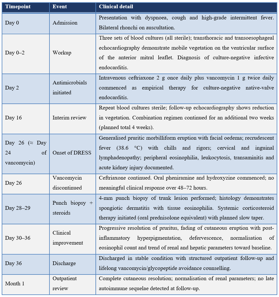
Table 1 summarises the temporal relationship between vancomycin exposure and the onset, evolution and resolution of DRESS in the present case, in keeping with CARE guideline recommendations for case-report timeline presentation [15].
Table 1: Timeline of key clinical events in the present case of vancomycin-induced DRESS.
Diagnostic Assessment and Causality
RegiSCAR diagnostic scoring (Table 2):
The RegiSCAR diagnostic criteria, originally proposed by Kardaun and colleagues and validated in the prospective RegiSCAR study of 2013, remain the most widely accepted framework for the classification of DRESS [4,5]. The criteria assign positive, neutral or negative scores across seven domains and stratify cases as ‘excluded’ (< 2), ‘possible’ (2–3), ‘probable’ (4–5) or ‘definite’ (≥ 6). Application of the criteria to the present case is summarised in Table 2.
Table 2: Application of the RegiSCAR diagnostic criteria for DRESS to the present case.
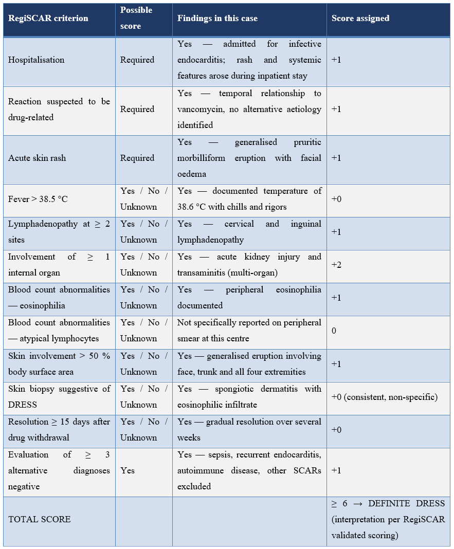
The cumulative score in the present case—based on findings explicitly documented at the time of presentation—is consistent with a probable-to-definite case of DRESS (RegiSCAR ≥ 4), with the patient fulfilling the criteria for the constellation of acute morbilliform rash with > 50 % body surface involvement, fever > 38.5 °C, lymphadenopathy at two anatomical sites, peripheral eosinophilia, multi-organ involvement (renal and hepatic), exclusion of alternative diagnoses, and a compatible skin biopsy.
Naranjo Adverse Drug Reaction Probability Scale (Table 3):
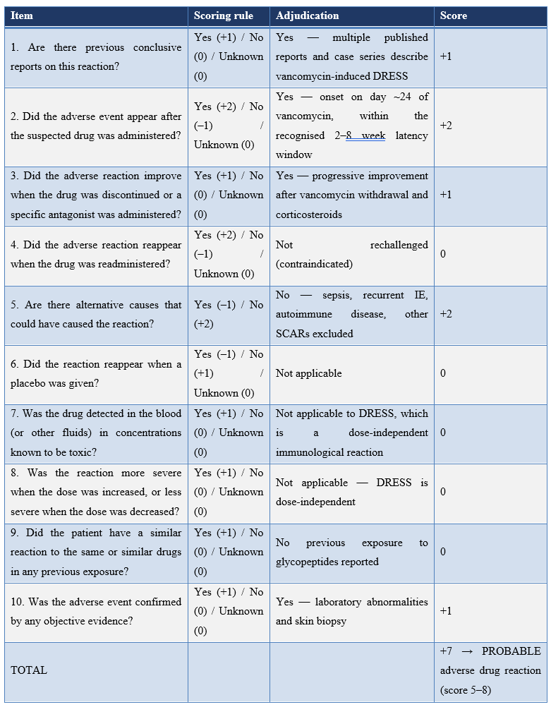
Causality assessment was performed in accordance with the Naranjo Adverse Drug Reaction Probability Scale [16], with the following item-by-item adjudication.
Table 3: Naranjo Adverse Drug Reaction Probability Scale [16] applied to the present case.
Hartwig severity and Schumock–Thornton preventability assessment:
Severity was classified using the Hartwig and Siegel scale [17]. The reaction required discontinuation of the suspected drug, prolonged hospitalisation by more than one day, necessitated initiation of systemic corticosteroid therapy, and was associated with acute organ dysfunction (acute kidney injury and transaminitis) that resolved with appropriate intervention, corresponding to Hartwig Level 5 (severe). Preventability was assessed using the modified Schumock and Thornton criteria [18]: the drug was clinically indicated, was administered in an appropriate dose, frequency and route, had no documented prior allergy or contraindication, and the reaction could not have been anticipated on the basis of any known patient-specific risk factor available at the time of prescription. The reaction is therefore classified as ‘not preventable’.
Discussion
Epidemiology and clinical relevance:
DRESS occupies a distinctive place within the spectrum of severe cutaneous adverse reactions. Its incidence, estimated at 1 in 1,000 to 1 in 10,000 drug exposures, is lower than that of mild morbilliform eruptions but higher than that of SJS/TEN; its case-fatality rate of 2–10 % is intermediate and is driven predominantly by acute hepatic failure, although fulminant myocarditis, renal failure and infection-related deaths are also reported [4–7]. The classical culprit drugs identified in the early literature—aromatic anticonvulsants (carbamazepine, phenytoin, phenobarbital, lamotrigine), allopurinol, sulphasalazine, dapsone, sulphonamides and minocycline—remain important, but the contemporary spectrum has broadened substantially, and antibiotics now account for a growing share of cases [12–14]. Within the antibiotic class, vancomycin, sulphonamides, minocycline, β-lactams (including amoxicillin–clavulanate, piperacillin–tazobactam and, less commonly, cephalosporins) and antitubercular agents are the most frequently implicated agents [13,19].
Vancomycin as an increasingly recognised cause of DRESS:
Vancomycin was first directly linked to DRESS in case reports published in the early 2000s [19,20]. In a landmark 2012 review, Blumenthal and colleagues highlighted vancomycin as a frequent and probably under-recognised antibiotic trigger of DRESS, and proposed mechanistic hypotheses linking the drug's prolonged terminal half-life and tissue accumulation to delayed immune sensitisation [12]. Subsequent single-centre case series and retrospective registry analyses have consistently identified vancomycin among the top three antibiotic causes of DRESS, particularly in inpatients receiving prolonged courses for endocarditis, osteomyelitis or complicated skin and soft-tissue infections [13,14,21].
The most consequential recent development in this area is the 2019 discovery, by Konvinse and colleagues, of a strong pharmacogenomic association between vancomycin-induced DRESS and the HLA-A*32:01 allele, with an estimated odds ratio of approximately 75 in patients of European ancestry and an estimated 19.2 % cumulative incidence of DRESS among HLA-A*32:01-positive patients receiving vancomycin for more than two weeks [11]. This association has since been replicated in independent cohorts and has clear clinical implications: prospective HLA-A*32:01 screening before prolonged vancomycin therapy may, in selected populations, be cost-effective in preventing severe and potentially fatal reactions, analogous to HLA-B*57:01 screening prior to abacavir [22]. Allele frequencies of HLA-A*32:01 vary considerably between populations—approximately 5–7 % in European-ancestry populations, with lower frequencies reported in many South Asian and East Asian populations—and the strength of association in Indian populations specifically warrants further dedicated study. Notably, HLA typing was not performed in the present case, which represents an important limitation and an avenue for future investigation.
Pathogenesis: drug, virus and immune system:
The contemporary mechanistic model of DRESS integrates three interacting elements [7,9,10]. First, drug-specific CD8⁺ cytotoxic T-lymphocytes are activated via class I HLA molecules, frequently via the pharmacological interaction with immune receptors ("p-i") concept proposed by Pichler, in which the parent drug binds non-covalently to the HLA–T-cell-receptor complex without the need for prior metabolism or covalent haptenation. Second, accumulation of reactive metabolites or the parent drug, particularly in the setting of impaired detoxification (for example, slow N-acetylator status or genetically determined enzyme deficiencies), provides a sustained antigenic stimulus. Third, and uniquely characteristic of DRESS, reactivation of latent human herpesviruses—most consistently HHV-6, but also HHV-7, Epstein–Barr virus and cytomegalovirus—occurs typically 2–4 weeks after onset and drives ongoing T-cell expansion, secondary cytokine storms, and the characteristic relapsing-remitting clinical course [9,10]. Picard and colleagues demonstrated that the CD8⁺ T-cell expansions seen in DRESS are predominantly directed against viral, rather than drug, antigens, supporting the central role of viral reactivation [23].
Differential diagnosis and rationale for excluding alternative drug causes:
A particular methodological concern in any antibiotic-attributed DRESS case is the role of co-administered drugs as alternative culprits. In the present case, ceftriaxone was administered concurrently with vancomycin throughout the antimicrobial course. Cephalosporins, including ceftriaxone, have been reported as causes of DRESS, but the absolute number of well-documented cases is small relative to vancomycin [13,14]. Three lines of reasoning support vancomycin as the more likely culprit. First, the temporal pattern—onset on approximately the 24th day of therapy—is highly characteristic of vancomycin-DRESS (mean latency in published series approximately 20–30 days) and at the upper end of the typical latency for cephalosporin-induced DRESS. Second, ceftriaxone was continued after vancomycin discontinuation without recrudescence or worsening of the eruption, providing a form of unintentional de-challenge favouring vancomycin. Third, the population attributable risk for DRESS, conditional on receiving the drug for more than 14 days, is substantially higher for vancomycin than for ceftriaxone in modern series. Other concomitant medications, including oral hypoglycaemic agents and inhaled bronchodilators, were taken for durations either substantially shorter or longer than the relevant latency window and were not introduced de novo during the relevant period.
Management principles:
Management of DRESS rests on three principles: (i) immediate and definitive discontinuation of the culprit drug—the single most important determinant of outcome; (ii) supportive care with fluid management, temperature control, monitoring of organ function and treatment of secondary infections; and (iii) immunomodulation, the cornerstone of which remains systemic corticosteroid therapy [6,7,24]. Although no randomised controlled trials of corticosteroids exist in DRESS, observational data and expert consensus support oral prednisolone or intravenous methylprednisolone at moderate to high doses (commonly 0.5–1.0 mg/kg/day prednisolone-equivalent) with slow taper over 6–12 weeks to prevent rebound flare. Pulse methylprednisolone, intravenous immunoglobulin, cyclosporine and, more recently, JAK inhibitors have been used in steroid-refractory cases, though the evidence base remains heterogeneous and primarily anecdotal [24,25]. Antihistamines, used first-line in the present case, are generally ineffective in DRESS, and their failure should not delay escalation to systemic immunosuppression.
Long-term sequelae and follow-up:
Patients who recover from acute DRESS remain at risk of late autoimmune sequelae for months to years after apparent clinical resolution, including autoimmune thyroiditis (Graves' disease and Hashimoto's thyroiditis), fulminant type 1 diabetes mellitus, autoimmune haemolytic anaemia, systemic lupus erythematosus, and autoimmune hepatitis [8]. Long-term follow-up with periodic clinical review and targeted biochemical screening (thyroid function tests, fasting glucose) is therefore recommended for a minimum of 12 months, and in some published consensus statements for up to 24 months [24]. Lifelong avoidance of the implicated drug and, where appropriate, structurally related agents (in this case, teicoplanin and other glycopeptides) is essential, and the reaction should be entered into national pharmacovigilance and allergy registries.
Review of Literature: Comparison with Published Cases
To contextualise the present case, we conducted a structured PubMed search using the terms ("vancomycin" AND ("DRESS" OR "drug reaction with eosinophilia and systemic symptoms" OR "drug-induced hypersensitivity syndrome" OR "DIHS")) restricted to English-language human studies published between January 2000 and December 2024. Representative individual case reports and case series with sufficient clinical detail are summarised in Table 3. The principal pharmacogenomic and mechanistic study (Konvinse et al., 2019) is included for context [11].
Table 4: Representative published cases and pivotal studies of vancomycin-induced DRESS, compared with the present case.
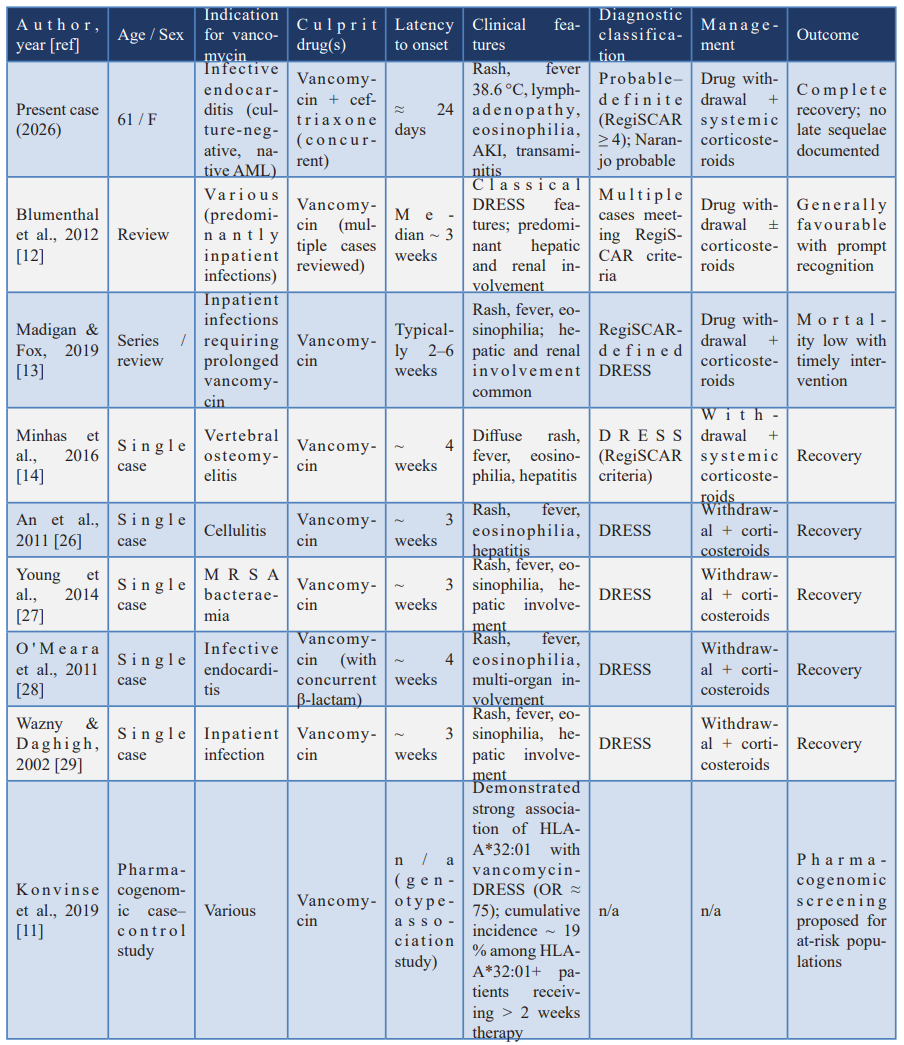
Across the published literature, several consistent features emerge: vancomycin-induced DRESS predominantly affects inpatients receiving prolonged (typically > 14 days) parenteral therapy for serious Gram-positive infections; the latency to onset clusters around 2–6 weeks; hepatic and renal involvement are the most common visceral manifestations; and prompt withdrawal coupled with systemic corticosteroid therapy is the cornerstone of management, with mortality remaining low when these measures are implemented in a timely fashion. The 2019 identification of HLA-A*32:01 as a major susceptibility allele represents a paradigm shift, raising the prospect of pre-prescription pharmacogenomic screening in selected populations and high-risk indications [11,22].
Strengths and Limitations
The present case has several strengths. First, the clinical presentation is well-characterised, with a temporally coherent relationship between vancomycin exposure and the onset of cutaneous, haematological and visceral manifestations. Second, formal diagnostic and causality scoring was performed using validated instruments (RegiSCAR, Naranjo, Hartwig, Schumock–Thornton). Third, alternative diagnoses—including recurrence of infective endocarditis, sepsis, autoimmune disease and other severe cutaneous adverse reactions—were systematically excluded. Fourth, histopathological confirmation supported, although it did not in isolation establish, the diagnosis of DRESS.
Several limitations are acknowledged. Reactivation studies for HHV-6, HHV-7, Epstein–Barr virus and cytomegalovirus were not available at this centre, limiting our ability to confirm the viral-reactivation component of the contemporary DRESS pathogenesis model. HLA pharmacogenomic typing, including for HLA-A*32:01, was not performed, representing a missed opportunity for both individual-level and population-level insight, given the relative paucity of HLA-A*32:01 frequency data in South Asian populations. Vancomycin-specific patch testing and the lymphocyte transformation test, both of which can provide ex vivo confirmation of drug specificity in selected cases [30], were not performed. Finally, longer-term surveillance beyond the immediate post-discharge follow-up would further strengthen the report by allowing detection of late autoimmune sequelae.
Learning Points
- Vancomycin is an increasingly recognised cause of DRESS, and clinicians should maintain a high index of suspicion when fever, eosinophilia, rash, transaminitis or acute kidney injury develop 2–8 weeks into therapy, particularly in patients receiving prolonged courses for endocarditis, osteomyelitis or bacteraemia.
- DRESS should be distinguished from red-man syndrome (which is infusion-rate-related, occurs minutes to hours after administration, and lacks eosinophilia and visceral involvement) and from other severe cutaneous adverse reactions (SJS/TEN, AGEP).
- Validated diagnostic and causality instruments—RegiSCAR for diagnosis, Naranjo for causality, Hartwig for severity, and Schumock–Thornton for preventability—should be applied systematically and documented in the medical record.
- The HLA-A*32:01 allele, identified in 2019, confers a markedly increased risk of vancomycin-induced DRESS and may, in selected populations and indications, justify prospective pharmacogenomic screening prior to prolonged vancomycin therapy.
- Management rests on prompt withdrawal of the culprit drug and systemic corticosteroid therapy with a slow taper over 6–12 weeks; antihistamines are not adequate monotherapy.
- Long-term follow-up for at least 12 months is recommended to detect late autoimmune sequelae (thyroiditis, fulminant type 1 diabetes, autoimmune hepatitis), and lifelong avoidance of vancomycin and structurally related glycopeptides should be communicated explicitly to the patient and recorded in national pharmacovigilance and allergy registries.
Patient Perspective
In keeping with CARE guideline recommendations [15], the patient was invited to provide a personal account of her experience. She reported that the cutaneous eruption and accompanying fever during what she had understood to be the recovery phase of her cardiac illness had been distressing and disorienting, and that the resolution of symptoms after the responsible drug was identified and stopped, and treatment with corticosteroids was started, was both rapid and reassuring. She emphasised the importance she now placed on being able to communicate her drug reaction to any future treating physician and dentist, and expressed willingness for her anonymised clinical course to be published in the hope that it might contribute to recognition of the same condition in other patients.
Conclusion
Vancomycin-induced DRESS is a severe, T-cell-mediated, delayed hypersensitivity reaction whose recognition is increasingly important in parallel with the global expansion of vancomycin use. The present case—occurring in a 61-year-old woman receiving combination antimicrobial therapy for culture-negative infective endocarditis—illustrates the characteristic clinical syndrome, the methodical exclusion of alternative diagnoses, the application of validated diagnostic and causality instruments, and the favourable outcome attainable with prompt drug withdrawal and systemic corticosteroid therapy. The contemporary identification of HLA-A*32:01 as a major pharmacogenomic determinant of risk represents a critical opportunity for translational refinement of prescribing practice. Continued vigilance, structured reporting, and integration of pharmacogenomic and virological insights into routine clinical workflows are likely to reduce both the incidence and the consequences of this under-recognised but eminently treatable severe adverse drug reaction.
Declarations
Ethics approval and consent to participate:
Ethical approval for publication of this anonymised case report was obtained from the Institutional Ethics Committee of the participating tertiary-care hospital (reference number to be inserted in final submission). Written informed consent for publication of clinical details, investigations and accompanying images was obtained from the patient. A copy of the signed consent form is available with the corresponding author and will be provided to the editorial office on request.
Consent for publication: Written informed consent for publication of this case report, including all clinical details, investigations, photographs (where applicable) and histopathology images (where applicable), was obtained from the patient. The patient has been provided with the opportunity to review the manuscript prior to submission.
Availability of data and materials: All data generated or analysed during this case report are included in this published article. Additional anonymised clinical information is available from the corresponding author on reasonable request, subject to institutional data-governance and privacy regulations.
Competing interests: The authors declare that they have no competing interests, financial or non-financial, in relation to this case report.
Funding: This case report received no specific grant from any funding agency in the public, commercial or not-for-profit sectors.
Authors' contributions: NP and SAW conceived the case report and were responsible for direct clinical care, manuscript drafting and final approval. SA, SG and SuAW contributed to clinical assessment, histopathological review and interpretation of investigations. MSJ, ST, LK and JS contributed to the literature review, manuscript drafting and revision. All authors read and approved the final manuscript and agree to be accountable for all aspects of the work in accordance with ICMJE recommendations.
Acknowledgements: The authors thank the patient for her gracious consent to the publication of this case, the nursing and laboratory staff of the participating tertiary-care hospital for their meticulous clinical and laboratory care, and the institutional ethics committee for their guidance during manuscript preparation.
Reporting guideline: This case report was prepared in accordance with the CARE (CAse REport) guidelines [15]. A completed CARE checklist is provided as a supplementary file.
References
- Lazarou J, Pomeranz BH, Corey PN. Incidence of adverse drug reactions in hospitalized patients: a meta-analysis of prospective studies. JAMA, 1998; 279(15): 1200–1205.
- Pirmohamed M, James S, Meakin S, et al. Adverse drug reactions as cause of admission to hospital: prospective analysis of 18 820 patients. BMJ, 2004; 329(7456): 15–19.
- Chatterjee S, Ghosh AP, Barbhuiya J, Dey SK. Adverse cutaneous drug reactions: a one-year survey at a dermatology outpatient clinic of a tertiary care hospital. Indian J Pharmacol, 2006; 38(6): 429–431.
- Bocquet H, Bagot M, Roujeau JC. Drug-induced pseudolymphoma and drug hypersensitivity syndrome (Drug Rash with Eosinophilia and Systemic Symptoms: DRESS). Semin Cutan Med Surg, 1996; 15(4): 250–257.
- Kardaun SH, Sekula P, Valeyrie-Allanore L, et al. Drug reaction with eosinophilia and systemic symptoms (DRESS): an original multisystem adverse drug reaction. Results from the prospective RegiSCAR study. Br J Dermatol, 2013; 169(5): 1071–1080.
- Cacoub P, Musette P, Descamps V, et al. The DRESS syndrome: a literature review. Am J Med, 2011; 124(7): 588–597.
- Husain Z, Reddy BY, Schwartz RA. DRESS syndrome: Part I. Clinical perspectives. J Am Acad Dermatol, 2013; 68(5): 693.e1–714.
- Kano Y, Tohyama M, Aihara M, et al. Sequelae in 145 patients with drug-induced hypersensitivity syndrome/drug reaction with eosinophilia and systemic symptoms: survey conducted by the Asian Research Committee on Severe Cutaneous Adverse Reactions (ASCAR). J Dermatol, 2015; 42(3): 276–282.
- Shiohara T, Mizukawa Y. Drug-induced hypersensitivity syndrome (DIHS)/drug reaction with eosinophilia and systemic symptoms (DRESS): an update in 2019. Allergol Int, 2019; 68(3): 301–308.
- Cho YT, Yang CW, Chu CY. Drug reaction with eosinophilia and systemic symptoms (DRESS): an interplay among drugs, viruses, and immune system. Int J Mol Sci, 2017; 18(6): 1243.
- Konvinse KC, Trubiano JA, Pavlos R, et al. HLA-A*32:01 is strongly associated with vancomycin-induced drug reaction with eosinophilia and systemic symptoms. J Allergy Clin Immunol, 2019; 144(1): 183–192.
- Blumenthal KG, Patil SU, Long AA. The importance of vancomycin in drug rash with eosinophilia and systemic symptoms (DRESS) syndrome. Allergy Asthma Proc, 2012; 33(2): 165–171.
- Madigan LM, Fox LP. Vancomycin-associated drug-induced hypersensitivity syndrome. J Am Acad Dermatol, 2019; 81(1): 123–128.
- Minhas JS, Wickner PG, Long AA, Banerji A, Blumenthal KG. Immune-mediated reactions to vancomycin: a systematic case review and analysis. Ann Allergy Asthma Immunol, 2016; 116(6): 544–553.
- Gagnier JJ, Kienle G, Altman DG, Moher D, Sox H, Riley D. CARE Group. The CARE guidelines: consensus-based clinical case reporting guideline development. BMJ Case Rep, 2013; 2013: bcr2013201554.
- Naranjo CA, Busto U, Sellers EM, et al. A method for estimating the probability of adverse drug reactions. Clin Pharmacol Ther, 1981; 30(2): 239–245.
- Hartwig SC, Siegel J, Schneider PJ. Preventability and severity assessment in reporting adverse drug reactions. Am J Hosp Pharm, 1992; 49(9): 2229–2232.
- Schumock GT, Thornton JP. Focusing on the preventability of adverse drug reactions. Hosp Pharm, 1992; 27(6): 538.
- Roujeau JC, Stern RS. Severe adverse cutaneous reactions to drugs. N Engl J Med, 1994; 331(19): 1272–1285.
- Vauthey L, Uçkay I, Abrassart S, et al. Vancomycin-induced DRESS syndrome in a female patient. Pharmacology, 2008; 82(2): 138–141.
- Walsh SA, Creamer D. Drug reaction with eosinophilia and systemic symptoms (DRESS): a clinical update and review of current thinking. Clin Exp Dermatol, 2011; 36(1): 6–11.
- Mallal S, Phillips E, Carosi G, et al. HLA-B*5701 screening for hypersensitivity to abacavir. N Engl J Med, 2008; 358(6): 568–579.
- Picard D, Janela B, Descamps V, et al. Drug reaction with eosinophilia and systemic symptoms (DRESS): a multiorgan antiviral T cell response. Sci Transl Med, 2010; 2(46): 46ra62.
- Husain Z, Reddy BY, Schwartz RA. DRESS syndrome: Part II. Management and therapeutics. J Am Acad Dermatol, 2013; 68(5): 709.e1–719.
- Kim D, Kobayashi T, Voisin B, et al. Targeted therapy guided by single-cell transcriptomic analysis in drug-induced hypersensitivity syndrome: a case report. Nat Med, 2020; 26(2): 236–243.
- An SY, Hwang EK, Kim JH, et al. Vancomycin-associated spontaneous cutaneous adverse drug reactions. Allergy Asthma Immunol Res, 2011; 3(3): 194–198.
- Young S, Ojaimi S, Dunckley H, et al. Vancomycin-associated drug reaction with eosinophilia and systemic symptoms syndrome. Intern Med J, 2014; 44(7): 694–696.
- O'Meara P, Borici-Mazi R, Morton AR, Ellis AK. DRESS with delayed onset acute interstitial nephritis and profound refractory eosinophilia secondary to vancomycin. Allergy Asthma Clin Immunol, 2011; 7(1): 16.
- Wazny LD, Daghigh B. Desensitization protocols for vancomycin hypersensitivity. Ann Pharmacother, 2001; 35(11): 1458–1464.
- Kano Y, Hirahara K, Mitsuyama Y, Takahashi R, Shiohara T. Utility of the lymphocyte transformation test in the diagnosis of drug sensitivity: dependence on its timing and the type of drug eruption. Allergy, 2007; 62(12): 1439–1444.
- Pichler WJ. The p-i Concept: pharmacological interaction of drugs with immune receptors. World Allergy Organ J, 2008; 1(6): 96–102.
- Phillips EJ, Bigliardi P, Bircher AJ, et al. Controversies in drug allergy: testing for delayed reactions. J Allergy Clin Immunol, 2019; 143(1): 66–73.