
Email id: info@ijclinmedcasereports.com
Pulmonary Decline After Radiotherapy for Cardiac Sarcoma: A Triad of Pneumonitis, Pulmonary Oedema, and Infection Presenting a Diagnostic and Therapeutic Challenge
Lau A1, Lim CH1,*, Williams M2 and Lane N1,3
1Respiratory Medicine, The Royal Victoria Infirmary, Newcastle upon Tyne, UK
2Cardiology Department, The Royal Victoria Infirmary, Newcastle upon Tyne, UK
3Translational and Clinical Research Institute, Newcastle University, Newcastle upon Tyne, UK
Received Date: 29/06/2026; Published Date: 02/09/2026
*Corresponding author: Cheng Hong Lim, Respiratory Medicine, The Royal Victoria Infirmary, Newcastle upon Tyne, UK
Abstract
Primary cardiac sarcomas are rare, aggressive malignancies, with leiomyosarcoma (LMS) representing an unusual subtype. Radiotherapy (RT) is often employed for palliation in unresectable cardiac tumours; However, the respiratory complications of this approach are not well documented. We report the case of a 66-year-old man with primary cardiac LMS involving the right atrium and inferior vena cava, treated with palliative RT, who subsequently developed hypoxia and bilateral pulmonary infiltrates extending beyond the irradiated field.
This presentation created a complex diagnostic challenge, with differential considerations including Radiation Pneumonitis (RP), pulmonary oedema, and infection. Despite treatment with corticosteroids, diuretics, and broad-spectrum antibiotics, his condition deteriorated, culminating in acute respiratory distress syndrome.
This case highlights the rarity of RP following RT for cardiac tumours, particularly when affecting non-irradiated lung, and it illustrates the diagnostic uncertainty that arises when cardiac and respiratory pathologies coexist. It emphasises the value of early post-treatment follow-up, careful use of imaging, and close multidisciplinary collaboration across specialities. Finally, it underscores the importance of clear and compassionate communication with patients and families when the prognosis is uncertain and clinical decline is rapid.
Introduction
Primary cardiac neoplasms are incredibly rare, with a reported lifetime incidence of ≤0.02% [1]. In contrast, secondary (metastatic) involvement of the heart is substantially more common [2]. Of the primary cardiac tumours, fewer than 10% are malignant; of those that are, the most common are classified histologically as sarcomas or lymphomas [3], with an overall incidence of ≤0.005% [4].
Among cardiac sarcomas, leiomyosarcomas (LMSs) account for approximately 10%. These typically arise in the left atrium and present between the fourth and sixth decades of life, with no observed sex predilection [3]. Unfortunately, malignant primary cardiac tumours carry a poor prognosis, with a mean survival of approximately six months following diagnosis [5].
Radiotherapy (RT), whether delivered with curative or palliative intent, can cause adverse effects to adjacent critical structures, despite significant advancements in radiation delivery techniques [6]. Radiation Pneumonitis (RP) is a common acute manifestation of radiation-induced lung injury (RILI) that typically presents between one and three months following the completion of radiotherapy. Conversely, radiation-induced pulmonary fibrosis represents a late sequela, usually developing between 6 to 24 months post-treatment [7].
The incidence of RP varies widely, affecting approximately 5-25 % of patients treated with thoracic irradiation [8]. Severity can be assessed with the five-grade Kong scale with stepwise treatment recommendations based on severity [9,10].
Risk factors contributing to the development of RP include mean lung radiation dose measured in gray (Gy) [11], non-smokers who lack the protective effect of tobacco, pre-existing lung conditions, and advanced patient age [12].
Case Presentation
We report the case of a 66-year-old White British male with a known diagnosis of an inter-atrial LMS, initially identified during a prior admission in April 2025 for decompensated heart failure refractory to diuretics. A cardiac biopsy confirmed LMS, which was Murine Double Minute 2 (MDM2) non-amplified. Because MDM2 amplification is characteristic of liposarcoma, its absence supports the diagnosis of LMS over liposarcoma. The tumour was deemed unresectable because of its infiltration into the right atrium and inferior vena cava as shown in imaging in Figures 1, 2 and 3. He was also unsuitable for stereotactic ablative body radiotherapy because of the tumour size and was not a candidate for systemic chemotherapy given his underlying heart failure and impaired renal function.
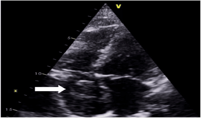
Figure 1: Echocardiogram demonstrating a large echo-dense mass (white arrow) in the right atrium, appearing to almost fill the chamber.
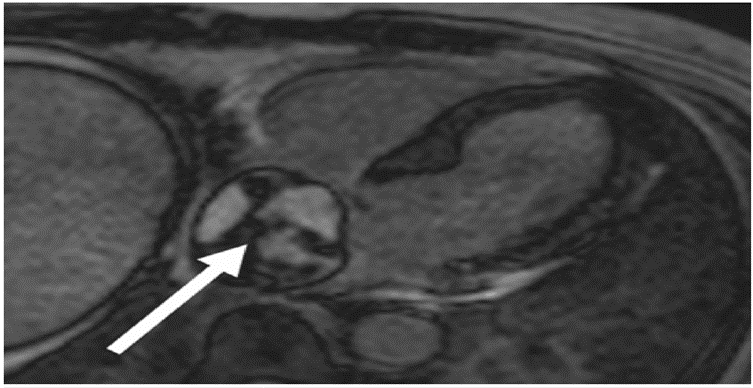
Figure 2: Cardiac MRI showing a large mass in the right atrium (white arrow) demonstrating heterogenous late gadolinium enhancement in keeping with a tumour.
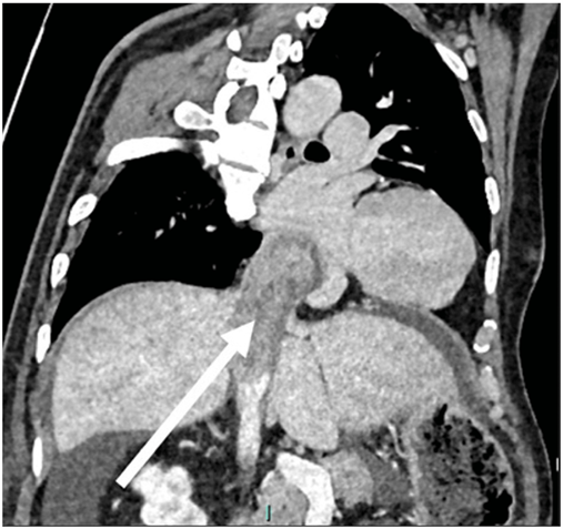
Figure 3: CT chest, abdomen and pelvis with contrast demonstrating a 10cm heterogeneously enhancing solid mass (white arrow) in the right atrium extending into the suprahepatic IVC.
A pre-radiotherapy staging computed tomography (CT) scan (Figure 4) demonstrated no evidence of metastatic disease. Additional findings included widespread bronchiectasis, with no evidence of focal pulmonary consolidation or pleural effusion.
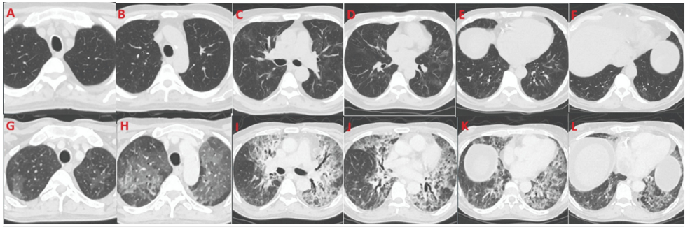
Figure 4: Comparison of CTCAP imaging between pre-admission scans (11/06/2024; panels A-F) and admission scans (06/09/2024; panels G-L), demonstrating the interval changes described in the text.
At his oncology follow-up in early September, he was noted to be recovering well clinically. Liver function tests revealed a mild hyperbilirubinemia (46 µmol/L; normal range 0-21 µmol/L) with normal alanine transaminase (ALT; 13 U/L; normal range 0-40 µmol/L), attributed to hepatic congestion secondary to fluid retention. A contrast-enhanced magnetic resonance imaging (MRI) liver as shown in Figure 5, revealed an indeterminate lesion that was atypical for a metastasis or primary liver tumour. Additional imaging findings were consistent with congestive hepatopathy, including caudate and lateral segment hypertrophy, posterior right liver notching, gallbladder wall oedema, and upper abdominal ascites-all of which were considered likely secondary to IVC obstruction caused by the cardiac tumour.
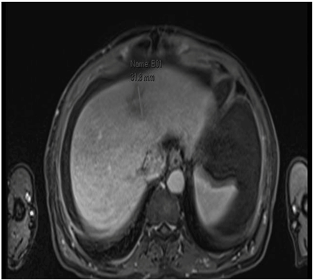
Figure 5: MRI Liver showing a 31.8 mm ill-defined area (labelled B01) of low intensity bordering segment 4A and 2 of the left lobe of the liver, best demonstrated on the portal venous phase series.
The management plan at the time was to repeat a CT Chest, Abdomen, and Pelvis (CTCAP) later in September to assess for disease progression. In the absence of metastatic disease, he would remain on active surveillance. Further palliative chemotherapy would be considered if there was radiological progression, contingent upon his functional status.
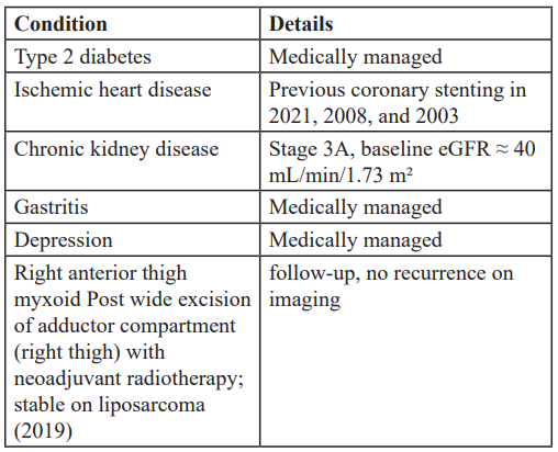
In the interim, the patient reported discomfort with swallowing, reduced oral intake, and poor appetite, which were attributed to oesophageal irritation following RT. He had been unable to continue furosemide after his supply was exhausted in the days preceding admission. On presentation, he reported a two-day history of progressive shortness of breath and dry cough, accompanied by worsening ascites and pleuritic left-sided chest pain since radiotherapy, exacerbated by deep inspiration and coughing. His past medical history is listed in Table 1. He was taking aspirin 75 mg once daily, bisoprolol 10 mg once daily, dapagliflozin 5 mg once daily, furosemide 40 mg twice daily, lansoprazole 15 mg once daily, and linagliptin 5 mg once daily. His nutritional intake was supported with Fortisip Compact twice daily. For cardiovascular symptoms, he was on isosorbide mononitrate 10 mg twice daily and used glyceryl trinitrate spray 400 micrograms per dose sublingually as required.
Initial Clinical Assessment
On admission, his National Early Warning Score (NEWS) [13] was 6 with a heart rate of 109 beats per minute and oxygen saturation of 96% while receiving 40% oxygen via a Venturi mask and tachypnoea; he was also apyrexial. Electrocardiography (ECG) as shown in Figure 6 demonstrated sinus tachycardia with right bundle branch block and no evidence of ischemia. His chest X-ray (CXR) (Figure 7) demonstrated bilateral mid-to-lower zone opacifications. The left mid-zone changes were felt to be most consistent with post-RT pneumonitis, given his recent exposure to high-dose thoracic irradiation, whereas the less well-defined right mid-zone opacity was considered to be possibly infective in nature, while accepting this could also represent RP.
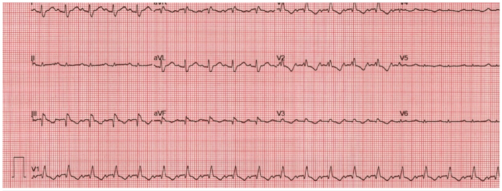
Figure 6: Admission electrocardiogram recorded at a paper speed of 25 mm/s and voltage calibration of 10 mm/mV, demonstrating a right bundle branch block pattern.
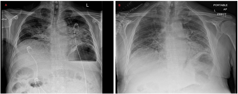
Figure 7: Serial chest radiographs demonstrating progression of pulmonary changes. (A) Admission chest radiograph showing bilateral mid-to-lower zone airspace opacification, more pronounced on the left, in keeping with early pneumonitic change or pulmonary oedema. (B) Repeat chest radiograph on Day 6 demonstrating persistent and relatively unchanged florid bilateral mid-to-lower zone consolidation with no pleural effusion, consistent with evolving severe pneumonitis.
Table 2: Blood results on admission compared to previous results after competing radiotherapy.
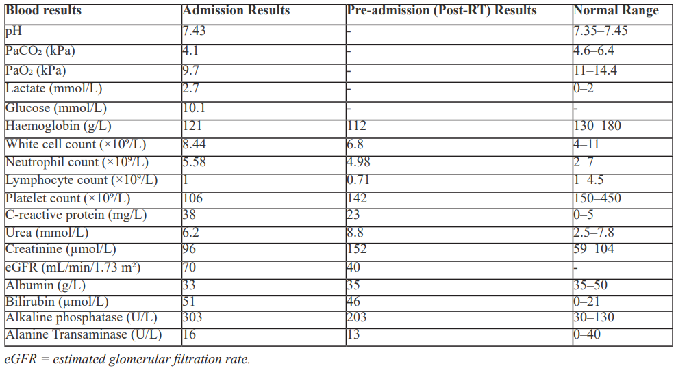
Admission blood tests (Table 2) revealed a mildly elevated Total White Cell count (TWC) and C-Reactive Protein (CRP) when compared to previous results. An arterial blood gas demonstrated type 1 respiratory failure. Respiratory viral swabs were negative for severe acute respiratory syndrome coronavirus 2, influenza virus, and respiratory syncytial virus.
The initial working diagnosis was pulmonary oedema, attributed to recent omission of furosemide, as well as ascites with deranged liver function tests and possible community-acquired pneumonia. Given his history and the initial differentials above, a cardiology opinion was sought. They felt the presentation was unlikely to be primarily cardiac in origin and more consistent with post-radiotherapy changes and ascites, because his echocardiogram demonstrated normal left ventricular ejection fraction (>55%) with normal RV function. A subsequent abdominal ultrasound (Figure 8), confirmed the presence of gross abdominal and pelvic ascites in keeping with congestive hepatopathy secondary to intrahepatic IVC obstruction.
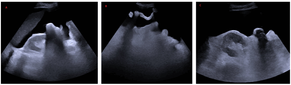
Figure 8: Admission abdominal ultrasound demonstrating gross ascites (A) Right upper quadrant view showing the liver and right kidney with a large volume of surrounding anechoic free fluid (B) Left iliac fossa view demonstrating marked free fluid tracking within the lower abdomen (C) Left kidney view with adjacent anechoic ascitic fluid confirming generalised intra-abdominal accumulation.
As his symptoms persisted and oxygen saturations continued to decline, he was escalated to IV piperacillin-tazobactam on Day 2 of admission. He was subsequently transferred to the Respiratory Support Unit and commenced on high-flow nasal cannula (HFNC) oxygen therapy.
A contrast-enhanced CTCAP, as shown in Figure 4, demonstrated ‘bilateral pneumonitic changes, with appearances highly suggestive of viral pneumonitis. The differential diagnoses include Acute Respiratory Distress Syndrome (ARDS) and pulmonary oedema.
On review, the patient’s dyspnoea and increasing oxygen requirements were considered multifactorial. The leading differential diagnoses included the following:
- Post-RT pneumonitis
- Cardiac dysfunction related to atrial tumour burden
- Superimposed lower respiratory tract infection
- Impaired diaphragmatic movement secondary to tense ascites
Other possibilities included aspiration pneumonia, pulmonary oedema from underlying heart failure, obstruction to blood flow by the right atrial mass, and less common causes such as lymphangitic spread or post-RT sequelae including cardiac dysfunction or pulmonary hypertension. A three-day empirical trial of dexamethasone 8 mg was commenced, and an ascitic tap was performed, which was negative for spontaneous bacterial peritonitis and demonstrated transudative fluid. Escalation to the critical care unit was not considered appropriate in view of his overall condition, particularly given concerns that invasive ventilation could exacerbate mechanical cardiac outflow obstruction from the sizeable right atrial tumour and underlying right heart failure.
His hypoxia was managed with HFNC (100% at 60 L/min) and continuous positive airway pressure (CPAP) at 8 cmH₂O, which was titrated in conjunction with physiotherapy support. Despite this, he experienced recurrent desaturation on minimal exertion and remained oxygen dependent.
At this stage, the patient was empirically treated with intravenous antibiotics, intravenous loop diuretics, and intravenous dexamethasone. This broad therapeutic approach was intended to address the range of differential diagnoses outlined. Despite these treatments, there was no clinical improvement and he continued to require 100% oxygen support via 60L/min HFNC and remained symptomatic with persistent dry cough, dyspnoea, and intermittent pyrexia. Therapeutic ascitic drainage provided only transient symptomatic relief. In parallel, the patient was commenced on insulin because of steroid-induced hyperglycaemia associated with the dexamethasone therapy.
After two days without clinical improvement on intravenous piperacillin-tazobactam, the antibiotic regimen was escalated to intravenous co-trimoxazole to provide coverage for potential opportunistic infections, particularly Pneumocystis jirovecii pneumonia (PJP), given the context of relative lymphopenia and recent thoracic radiotherapy. Further investigations were undertaken to explore infectious and inflammatory differentials; both Procalcitonin (PCT) and beta glucan were normal.
On day 6 of admission, a repeat CXR (Figure 7) demonstrated persistent florid bilateral mid-to-lower zone consolidative changes, with no significant radiological improvement.
Despite ongoing treatment, he continued to deteriorate and was unable to tolerate even brief breaks from CPAP, experiencing immediate desaturation even on HFNC at 60L/min and 100% FiO₂. The working impression was evolving towards refractory RP, with differential consideration of an atypical infection, notably PJP, in view of recent RT and relative immunosuppression
On day 8 of admission, discussions were ongoing with his family regarding the gravity of the situation and the lack of progress despite prolonged medical treatment. Despite broad-spectrum intravenous antibiotics, high-dose corticosteroids, and diuretics, there had been no clinical improvement. A PCT level suggested that bacterial infection was unlikely to be the primary driver of his condition.
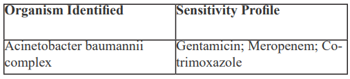
As a final therapeutic attempt, a three-day trial of intravenous (IV) methylprednisolone 250mg was commenced. On day 9, his antimicrobial coverage was escalated to include IV meropenem and clarithromycin to provide broader coverage for any occult or resistant infections. A sputum culture later grew A. baumannii complex, with sensitivities as summarised in Table 3.
Table 3: Sputum culture result showing A. baumannii complex and corresponding antimicrobial sensitivities.
Unfortunately, he did not demonstrate any further clinical improvement, and corticosteroid therapy was de-escalated. He became increasingly tachypnoeic and distressed, unable to tolerate CPAP, so focus shifted toward palliative care. He passed away peacefully on day 13 of admission with the medical cause of death recorded as pneumonitis.
Discussion
This case is notable due to the extreme rarity and aggressive behaviour of primary cardiac LMS [14,15]. Sadly, LMS is frequently unresectable at the time of diagnosis [16] and has a mean survival time of 17.5 months with treatment [17] and 6 months without treatment [18].
In contrast, atrial myxomas - the most common primary cardiac tumour - typically originate in the left atrium [19], are slow-growing, often curable with surgery, and associated with better outcomes [20,21,22]. While LMS most commonly involve the left atrium, our patient’s tumour involved the right atrium and extended into the IVC, a particularly rare presentation, previously reported only in isolated case studies such as this [23].
Consistent with existing literature, this tumour remained clinically silent until it led to symptoms related to mechanical obstruction and decompensated heart failure [16,23], prompting the initial diagnosis and later contributing to both admission and death. In this case, heart failure symptoms proved refractory even to high-dose IV diuretics and underscores the diagnostic challenges and rapid clinical deterioration typically associated with cardiac sarcomas - though RP was ultimately the driving process.
Data specifically addressing RP following RT for cardiac tumours are extremely limited. The majority of existing literature reports RP pertaining to patients treated with RT for thoracic malignancies such as lung, breast, and oesophageal cancers as well as lymphoma and in paediatric tumours [24-27].
Adjuvant RT has occasionally been employed for local control in cardiac sarcoma, but outcome data remain scarce. In a large modern series [28], postoperative therapy outcomes for primary cardiac sarcomas demonstrated the feasibility of adjuvant RT and improved local control, yet without reporting pulmonary toxicity. This absence of toxicity data highlights both the rarity of such cases and the likelihood of under-recognised RILI in this context. Our patient’s course therefore represents one of few documented instances of clinically significant RP following cardiac-directed RT.
In conventional RT, a significant volume of surrounding healthy lung tissue may be inadvertently irradiated, thereby increasing the risk of RP [12]. Although newer RT modalities-such as intensity-modulated radiotherapy (IMRT), volumetric-modulated arc therapy (VMAT), and stereotactic body radiotherapy (SBRT)-have minimized lung injury and are generally better tolerated, major pulmonary toxicity can still occur, with rates reported as high as 20% in some series [29,30].
The severity and distribution of RP may be influenced by several factors, including the total lung dose, volume of irradiated lung, fractionation scheme, chemotherapy, and patient-specific risk factors such as age, smoking history, and underlying comorbidities [12]. RILI can extend beyond the irradiated volume, manifesting histologically as organising pneumonia [6], or widespread interstitial pneumonitis, including contralateral lung involvement, and may be refractory to corticosteroid therapy [31].
Our patient initially presented with Grade 2 RP which unfortunately progressed to Grade 5 during his admission. He had received 30-40 Gy of RT, a dose range that is less commonly associated with overt radiographic changes of RP - these are typically observed at doses exceeding 40 Gy [32]. However, it is well-documented that doses above 20 Gy can increase the risk of developing radiation-induced lung injury [33-35].
Although he was a non-smoker - thereby lacking the paradoxical "protective" effect of tobacco exposure or smoking-induced pulmonary damage, which may confer relative radio-resistance by creating non-functional airspaces and fibrotic tissue [12] - he had a background of bronchiectasis. This underlying lung disease, evident on pre-RT CTCAP imaging, may have contributed to increased symptomatic burden [36,37].
At 66 years old, he was below the commonly cited age threshold of 70 years associated with increased risk of severe RP [38]. However, his low serum albumin at presentation (33 g/L) and the development of out-of-field RP were consistent with factors linked to poorer survival outcomes [39].
RP represents an acute inflammatory response of the lung parenchyma to radiation exposure, as illustrated in Figure 9. When the contra-lateral or non-irradiated lung is involved - as in this case - an immune-mediated lymphocytic alveolitis has been proposed as the underlying mechanism [40]. Although CXR frequently demonstrate airspace opacities, these findings are non-specific as CT is more sensitive at identifying parenchymal abnormalities with ground-glass opacities (GGO) and airspace consolidation being the most commonly reported features [6,7,41,42], as evident in this case from the post-radiotherapy CT (Figure 4).
The diagnosis of RP is more straightforward when there is a known history of thoracic RT and a clear demarcation of radiographic changes correlating with the irradiation field. In atypical distributions, however, GGOs - defined as areas of increased pulmonary attenuation without obscuration of bronchial and vascular margins - are non-specific and can represent a broad differential, including infectious pneumonia, opportunistic infections such as PJP, chronic interstitial lung disease, and acute alveolar processes such as pulmonary oedema [7].
In this case, diagnostic interpretation was confounded by multiple overlapping factors: a history of heart failure with missed doses of diuretics upon presentation, recent RT targeted at an atrial mass (rather than lung tissue), and potential infectious aetiologies in an immunocompromised patient. Consequently, our patient was treated empirically for all three leading differential diagnoses - receiving IV diuretics, broad-spectrum antibiotics, and high-dose corticosteroids, the latter being the mainstay of therapy for RILI and acute RP [6,7,10].
This therapeutic approach was initiated with an awareness of the risks associated with systemic corticosteroids, including sodium and fluid retention [43], and worsening oedema - an adverse prognostic marker in patients with cardiogenic shock [44]. While some isolated reports suggest corticosteroids may improve congestion, neurohormonal status, and renal function in selected heart failure patients [45], our patient received a combination of corticosteroids with both glucocorticoid (e.g. dexamethasone) and significant mineralocorticoid activity (e.g. methylprednisolone, prednisolone) [46] in an effort to mitigate the severity of acute RP. Unfortunately, this strategy did not prevent progression to ARDS, a condition associated with high mortality [40,41].
Moreover, moderate to high doses of systemic corticosteroids increase susceptibility to infections, including life-threatening opportunistic pathogens [8,43,47]. This risk was compounded in our patient by active cardiac malignancy, recent RT, and advanced age - all factors associated with impaired host immunity and increased infection risk [48]. In light of this, he was empirically treated with broad-spectrum IV antibiotics throughout his admission, initially piperacillin-tazobactam followed by co-trimoxazole, and subsequently meropenem. While initial microbiological cultures were negative, a later sputum culture identified Acinetobacter baumannii complex, sensitive to both meropenem and co-trimoxazole. This finding supported the antimicrobial regimen, though the likelihood of A. baumannii being pathogenic or causative in this clinical context was considered low.
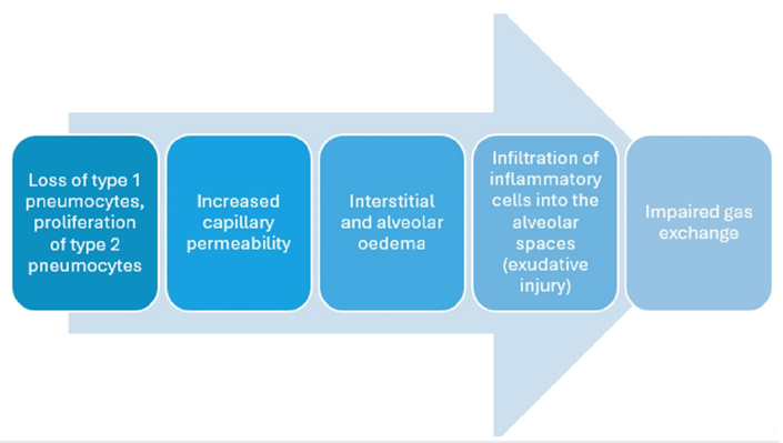
Figure 9: Author-generated diagram of the pathophysiology of radiation pneumonitis, informed by references [7,40].
Conclusion
This case illustrates the clinical challenges posed by a rare primary cardiac LMS involving the right atrium and IVC, complicated by severe RP. Diagnosis and management were particularly difficult, and the patient’s deterioration could not be reversed despite optimal treatment for the principle differential diagnoses: RP, pulmonary oedema from heart failure, and infection. Importantly, the case underlines the need to consider RP even outside the irradiated field, particularly when new respiratory symptoms emerge after radiotherapy.
MDT collaboration between cardiology, oncology, respiratory medicine, and radiology was vital in navigating this complex, multisystem presentation. Equally, this case highlights the importance of open, compassionate, and ongoing discussions with patients and families when prognosis is uncertain and treatment options are limited. Clear communication and shared decision-making remain central to patient-centred care, especially in rapidly evolving cancer scenarios.
Author Contributions: All authors have reviewed the final version to be published and agreed to be accountable for all aspects of the work.
Concept and design: Aaron Lau, Nicholas D. Lane, Cheng Hong Lim
Acquisition, analysis, or interpretation of data: Aaron Lau, Matthew Williams, Nicholas D. Lane, Cheng Hong Lim
Drafting of the manuscript: Aaron Lau, Nicholas D. Lane, Cheng Hong Lim
Critical review of the manuscript for important intellectual content: Aaron Lau, Matthew Williams, Nicholas D. Lane, Cheng Hong Lim
Supervision: Aaron Lau, Matthew Williams, Nicholas D. Lane, Cheng Hong Lim
Disclosure
Human subjects: Informed consent for treatment and open access publication was obtained or waived by all participants in this study. Royal Victoria Infirmary ethics committee issued approval -. Approval and consent obtained from family members and next of kin, verbally and written consent.
Conflicts of interest: In compliance with the ICMJE uniform disclosure form, all authors declare the following:
Payment/services info: All authors have declared that no financial support was received from any organization for the submitted work.
Financial relationships: All authors have declared that they have no financial relationships at present or within the previous three years with any organizations that might have an interest in the submitted work.
Other relationships: All authors have declared that there are no other relationships or activities that could appear to have influenced the submitted work.
References
- El Bardissi AW, Dearani JA, Daly RC, et al. Analysis of benign ventricular tumors: Long-term outcome after The Journal of Thoracic and Cardiovascular Surgery, 2008; 135: 1061-1068. doi: 10.1016/j.jtcvs.2007.10.048.
- Reynen K. Frequency of primary tumors of the heart . The American Journal of Cardiology, 1996; 77: 107. doi: 1016/S0002-9149(97)89149-7.
- Maleszewski J, Basso C, Bois M, et al. The 2021 WHO Classification of Tumors of the Heart . Journal of Thoracic Oncology, 2022; 17: 510-518. doi: 1016/j.jtho.2021.10.021.
- Molina J, Edwards J, Ward H. Primary Cardiac Tumors: Experience at the University of Minnesota . The Thoracic and Cardiovascular Surgeon, 1990; 38: 183-191. doi: 1055/s-2007-1014064.
- Neragi-Miandoab S, Kim J, Vlahakes GJ. Malignant Tumours of the Heart: A Review of Tumour Type, Diagnosis and Therapy. Clinical Oncology, 2007; 19: 748-756. doi: 1016/j.clon.2007.06.009.
- Benveniste MF, Gomez D, Carter BW, et al. Recognizing Radiation Therapy-related Complications in the RadioGraphics, 2019; 39: 344-366. doi: 10.1148/rg.2019180061
- Gaillard F, Campos A, Murphy A,
- Hanania AN, Mainwaring W, Ghebre YT, Hanania NA, Ludwig M. Radiation-Induced Lung Injury . Chest, 2019; 156: 150-162. doi: 1016/j.chest.2019.03.033.
- Kong FM, Hayman JA, Griffith KA, et al. Final toxicity results of a radiation-dose escalation study in patients with non-small-cell lung cancer (NSCLC): Predictors for radiation pneumonitis and fibrosis. International Journal of Radiation Oncology*Biology*Physics, 2006; 65: 1075-1086. doi: 1016/j.ijrobp.2006.01.051.
- Fendler W, Tomasik B, Atkins K, Stawiski K, Chałubińska‑Fendler J, Kozono D. The clinician’s guide to radiotherapy complications. Polish Archives of Internal Medicine, 2022; 28: 132. doi: 20452/pamw.16190.
- Kwa SLS, Lebesque JV, Theuws JCM, et al. Radiation pneumonitis as a function of mean lung dose: an analysis of pooled data of 540 patients. International Journal of Radiation Oncology*Biology*Physics, 1998; 42: 1-9. doi: 1016/S0360-3016(98)00196-5.
- Arroyo-Hernández M, Maldonado F, Lozano-Ruiz F, Muñoz-Montaño W, Nuñez-Baez M, Arrieta O. Radiation-induced lung injury: current evidence. BMC Pulmonary Medicine, 2021; 6: 9. doi: 1186/s12890-020-01376-4.
- Scott LJ, Redmond NM, Garrett J, Whiting P, Northstone K, Pullyblank A. Distributions of the National Early Warning Score (NEWS) across a healthcare system following a large-scale roll-out. Emergency Medicine Journal, 2019; 36: 287-292. doi: 1136/emermed-2018-208140.
- Janopaul-Naylor J, Kanter KR, Flampouri S, Nguyen V, Olson TA, Eaton BR. Adjuvant chemoradiation for high-grade cardiac leiomyosarcoma in a child: Case report and review of literature. Pediatric Blood & Cancer, 2021; 14: 68. doi: 1002/pbc.29241.
- Muthukumar L, Jan MF, Taylor R, Crouch J, Neitzel G, Tajik AJ. Biatrial High-Grade Leiomyosarcoma With Nine-Year Post-Surgical Survival. The Annals of Thoracic Surgery, 2017; 103: 43-45. doi: 1016/j.athoracsur.2016.06.063.
- Nakashima K, Inatsu H, Kitamura K. Primary Cardiac Leiomyosarcoma: A 27-Month Survival with Surgery and Chemotherapy. Internal Medicine, 2017; 56: 2145-2149. doi: 2169/internalmedicine.6965-15.
- Chen TWW, Loong HH, Srikanthan A, et al. Primary cardiac sarcomas: A multi-national retrospective Cancer medicine, 2019; 8: 104-110. doi: 10.1002/cam4.1897.
- Poole GV, Meredith JW, Breyer RH, Mills SA. Surgical Implications in Malignant Cardiac Disease. The Annals of Thoracic Surgery, 1983; 36: 484-491. doi: 1016/S0003-4975(10)60494-8.
- Gošev I, Paić F, Đurić Ž, Gošev M, Ivčević S, Jakuš FB, et al. Cardiac myxoma the great imitators: Comprehensive histopathological and molecular approach. International Journal of Cardiology, 2013; 164: 7. doi: 1016/j.ijcard.2011.12.052.
- DʼAlfonso A, Catania S, Pierri MD, et al. Atrial myxoma: a 25-year single-institutional follow-up study . Journal of Cardiovascular Medicine, 2008; 9: 178-181. doi: 2459/JCM.0b013e3281ac22cb.
- Elsherif Z, Mahmood N, Ahmed AM: 30-year follow-up of an unoperated left atrial myxoma: a case report . European heart journal. Case reports, 2020; 4: 1-5. doi: 1093/ehjcr/ytaa310.
- Jiang CX, Wang JG, Qi RD, et al. Long-term outcome of patients with atrial myxoma after surgical intervention: analysis of 403 cases. Journal of geriatric cardiology: JGC, 2019; 16: 338-343. doi: 11909/j.issn.1671-5411.2019.04.003.
- Parissis H, Akbar MT, Young V. Primary leiomyosarcoma of the right atrium: a case report and literature update. Journal of cardiothoracic surgery, 2010; 12: 80. doi: 1186/1749-8090-5-80.
- Lewis GD, Agrusa JE, Teh BS, Gramatges MM, Kothari V, Allen CE, et al. Radiation pneumonitis in pediatric Hodgkin lymphoma patients receiving radiation therapy to the chest. Practical Radiation Oncology, 2018; 8: 364-368. doi: 1016/j.prro.2018.01.007.
- Pinnix CC, Huo J, Milgrom SA, et al. Using benchmarked lung radiation dose constraints to predict pneumonitis risk: Developing a nomogram for patients with mediastinal lymphoma. Advances in Radiation Oncology, 2018; 3: 372-381. doi: 1016/j.adro.2018.03.005.
- Whelan TJ, Olivotto IA, Parulekar WR, et al. Regional Nodal Irradiation in Early-Stage Breast Cancer . New England Journal of Medicine, 2015; 23: 307-316. doi: 1056/NEJMoa1415340.
- Yamaguchi S, Ohguri T, Matsuki Y, et al. Radiotherapy for thoracic tumors: association between subclinical interstitial lung disease and fatal radiation pneumonitis. International Journal of Clinical Oncology, 2015; 11: 45-52. doi: 1007/s10147-014-0679-1.
- Wu Y, Million L, Moding EJ, Scott G, Berry M, Ganjoo KN. The impact of postoperative therapy on primary cardiac sarcoma. The Journal of Thoracic and Cardiovascular Surgery, 2018; 156: 2194-2203. doi: 1016/j.jtcvs.2018.04.127.
- Prezzano KM, Ma SJ, Hermann GM, Rivers CI, Gomez-Suescun JA, Singh AK. Stereotactic body radiation therapy for non-small cell lung cancer: A review. World Journal of Clinical Oncology, 2019; 10: 14-27. 5306/wjco.v10.i1.14.
- van Sörnsen de Koste J, Voet P, Dirkx M, van Meerbeeck J, Senan S. An evaluation of two techniques for beam intensity modulation in patients irradiated for stage III non-small cell lung cancer. Lung Cancer, 2001; 32: 145-153. doi: 1016/S0169-5002(00)00214-2.
- Keffer S, Guy CL, Weiss E. Fatal Radiation Pneumonitis: Literature Review and Case Series. Advances in Radiation Oncology, 2020; 5: 238-249. doi: 1016/j.adro.2019.08.010.
- Libshitz HI, Southard ME. Complications of radiation therapy: The thorax . Seminars in Roentgenology, 1974; 9: 41-49. doi: 1016/0037-198X(74)90008-X.
- Matsuo Y, Shibuya K, Nakamura M, et al. Dose-Volume Metrics Associated with Radiation Pneumonitis After Stereotactic Body Radiation Therapy for Lung Cancer. International Journal of Radiation Oncology*Biology*Physics, 2012; 83: 545-549. doi: 1016/j.ijrobp.2012.01.018.
- Tsujino K, Hirota S, Endo M, et al.: Predictive value of dose-volume histogram parameters for predicting radiation pneumonitis after concurrent chemoradiation for lung cancer. International Journal of Radiation Oncology*Biology*Physics, 2003; 55: 110-115. doi: 1016/S0360-3016(02)03807-5.
- Yorke ED, Jackson A, Rosenzweig KE, et al. Dose-volume factors contributing to the incidence of radiation pneumonitis in non-small-cell lung cancer patients treated with three-dimensional conformal radiation International Journal of Radiation Oncology*Biology*Physics, 2002; 54: 329-339. doi: 10.1016/S0360-3016(02)02929-2.
- Ren C, Ji T, Liu T, Dang J, Li G. The risk and predictors for severe radiation pneumonitis in lung cancer patients treated with thoracic reirradiation. Radiation Oncology, 2018; 16: 69. doi: 1186/s13014-018-1016-z.
- Sanuki N, Ono A, Komatsu E, et al. Association of Computed Tomography-detected Pulmonary Interstitial Changes with Severe Radiation Pneumonitis for Patients Treated with Thoracic Radiotherapy. Journal of Radiation Research, 2012; 53: 110-116. doi: 1269/jrr.110142.
- Jin H, Tucker SL, Liu HH, et al. Dose-volume thresholds and smoking status for the risk of treatment-related pneumonitis in inoperable non-small cell lung cancer treated with definitive radiotherapy. Radiotherapy and Oncology, 2009; 91: 427-432. doi: 1016/j.radonc.2008.09.009.
- Wang JY, Chen KY, Wang JT, et al. Outcome and prognostic factors for patients with non-small-cell lung cancer and severe radiation pneumonitis. International Journal of Radiation Oncology*Biology*Physics, 2002; 54: 735-741. doi: 1016/S0360-3016(02)02994-2.
- Hassaballa HA, Cohen ES, Khan AJ, Ali A, Bonomi P, Rubin DB. Positron Emission Tomography Demonstrates Radiation-Induced Changes to Nonirradiated Lungs in Lung Cancer Patients Treated with Radiation and Chemotherapy. Chest, 2005; 128: 1448-1452. doi: 1378/chest.128.3.1448.
- Choi YW, Munden RF, Erasmus JJ, Joo Park K, Chung WK, Jeon SC, et al. Effects of Radiation Therapy on the Lung: Radiologic Appearances and Differential Diagnosis. RadioGraphics, 2004; 24: 985-997. doi: 1148/rg.244035160.
- Ikezoe J, Takashima S, Morimoto S, et al. CT appearance of acute radiation-induced injury in the lung . American Journal of Roentgenology, 1981; 150: 765-770. doi: 2214/ajr.150.4.765.
- Yasir M, Goyal A, Sonthalia S. Corticosteroid Adverse Effect StatPearls [Internet] (ed): StatPearls Publishing, Treasure Island (FL), 2025.
- Fan HP, Zhou Y, Zhou Y, Jin J, Hu TY. Association between short-term systemic use of glucocorticoids and prognosis of cardiogenic shock: a retrospective analysis. BMC Anesthesiology, 2023; 23: 169. doi: 1186/s12871-023-02131-y.
- Massari F, Mastropasqua F, Iacoviello M, Nuzzolese V, Torres D, Parrinello G. The glucocorticoid in acute decompensated heart failure: Dr Jekyll or Mr Hyde? The American Journal of Emergency Medicine, 2012; 30: 517-515. doi: 1016/j.ajem.2011.01.023.
- Amabile CM, Spencer AP. Keeping Your Patient with Heart Failure Safe . Archives of Internal Medicine, 2004; 7: 709. doi: 1001/archinte.164.7.709
- Li FJ, Yuan X, Chen C, Zhu ML, Bai CQ, Wang RJ. Clinical Analysis of Severe Radiation Pneumonia. International Journal of General Medicine, 2021; 14: 2581-2588. doi: 2147/IJGM.S311569.
- Curtis JR, Patkar N, Xie A, et al. Risk of serious bacterial infections among rheumatoid arthritis patients exposed to tumor necrosis factor α antagonists. Arthritis Rheum, 2007; 56: 1125-1133. doi: 10.1002/art.22504.