Aneurysm of the Ascending Aorta, A Rare and Serious Complication of Behçet's Disease: About a Case
Bouamoud A*, Bouazaze M, Zahidi Alaoui H, Malki M and Pr Asfalou I
Mohamed V Military Instruction Hospital, Rabat, Morocco
Avicenna University Hospital Rabat, Morocco
Received Date: 08/05/2025; Published Date: 13/06/2025
*Corresponding author: Bouamoud A, Mohamed V Military Instruction Hospital, Rabat ; Avicenna University Hospital Rabat, Morocco
Abstract
Arterial involvement in Behçet's disease occurs in 2 to 12% of patients and results in obliterating lesions and/or aneurysmals predominant on the large trunks. Cardiac complications are rarer (1 to 6%) affecting all three tunics.
Ascending aortic aneurysm is a very rare complication of Behcet's disease, the few cases described in the literature were almost always associated with aortic insufficiency.
We report the observation of a 75-year-old patient followed for Behcet's disease, in whom the diagnosis of an asymptomatic ascending aortic aneurysm was retained by transthoracic echocardiography and CT scan, with the absence of aortic leakage, something that makes the particularity of our observation.
Through this clinical case, we discuss the epidemiological, histological, therapeutic and evolutionary particularity of this rare entity.
Keywords: Behcet; Aneurysm; Ascending thoracic aorta; Surgery
Introduction
Behçet's disease (MB) is a systemic vasculitis, described in 1937 by Behçet, with a mainly venous tropism. Arterial involvement is present in only 2 to 8% of cases. It has three aspects ; These can be stenosis, thrombosis or more often arterial aneurysms.
Since the publication of the first case of aortic aneurysm during Behcet's disease in 1961, the number of published cases has continued to increase [1].
Aneurysm of the ascending aorta is a very rare complication of Behçet's disease, the few cases described in the literature were almost always associated with aortic insufficiency. We report the case of an aneurysm of the ascending aorta in the context of behcet's disease, with the absence of an aortic leak, which makes the particularity of our case.
Clinical Case
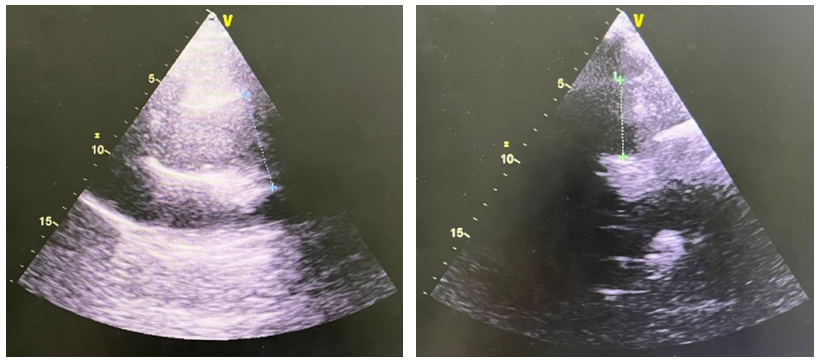
A 75-year-old patient, with cardiovascular risk factors for hypertension (hypertension) on dual therapy (ACE inhibitor and amlodipine), known to be a carrier of Behcet's disease on colchicine, asymptomatic patient, presented to the cardiology department for a control transthoracic echocardiography (ETT) as a follow-up assessment of the impact of his hypertension. ETT objectifies an aneurysm of the ascending thoracic aorta from the sinotubular junction measuring 59 mm with a normal-sized valsalva sinus and with no aortic leakage.
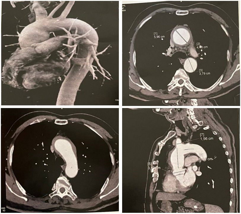
A cervico-thoracic CT angioCT showed an aneurysm of the ascending thoracic aorta measuring 59 mm in section, extended from the sinotubular junction to the level of the horizontal thoracic aorta, with circumferential thickening of an inflammatory nature.
The surgical indication was retained, however the patient refused any surgery.

Figure 1: Transthoracic echocardiographic images showing an ascending aortic aneurysm after the sinotubular junction sparing the first portions of the ascending aorta.

Figure 2: Cervicothoracic CT angiography images showing an aneurysmal dilation of the ascending thoracic aorta, measured at 59 mm in section, with homogeneous circumferential parietal thickening measured at 5.7 mm and a circulating lumen of 45 mm, originating from the sinotubular junction to the start of the brachiocephalic arterial trunk which measures 15.6 mm in section.
Discussion
Vascular involvement of Behcet's disease is common with predominance of men and can present in a wide variety of forms, 37% venous involvement, 12% arterial involvement, and 6% cardiac manifestations [1].
Arterial damage is currently better recognized and observed in 3% to 5% of cases depending on the series. This frequency is probably underestimated if we take into account autooptic data where arterial involvement is observed in one in three patients.
The time between diagnosis and the appearance of aneurysmal vascular involvement is variable; In the literature, this period varies from 18 to 53 months [2].
They are described as panvasculitis affecting small and large arteries with a possibility of aneurysmal and occlusive expression.
Aneurysms are the most common mode of arterial expression. They generally complicate a true canker sore and are the leading cause of death in Behçet's disease. They are often multifocal with a great predilection for the abdominal aorta, femoral and pulmonary arteries, so simultaneous aortic and pulmonary arterial or carotid arterial locations are not exceptional [2].
Aortic involvement is mainly represented by the aneurysm of the abdominal aorta, however the thoracic location was rarely described in the literature. The cases described in the literature mainly concern the descending thoracic aorta [3]. However, involvement of the ascending thoracic aorta is very rarely described and the few cases described were associated with severe aortic leakage [4-7] .
Histologically, it is vasa-vasorum vasority vasa devoid of giant cells leading to fragmentation and rupture of the media with deposition of immune complexes. These lesions lead either to the formation of a real aneurysm, or to the perforation responsible for a false aneurysm. In affected arteries, infiltrative lesions of the media and adventitia occur at the beginning and are subsequently followed by destructive and fibrous lesions of the media. In the active phase, granulomatous lesions similar to those observed during Takayasu's disease are frequently observed. Sacciform aneurysms are probably the result of severe destruction of the media by intense inflammatory reactions [2].
The direct role of vascular trauma is well known, illustrated by the occurrence of aneurysms at the arterial puncture site or at the suture point of vascular bypasses, performing a real pathergy test at the level of the artery. The role of tobacco overconsumption is highlighted in a few studies. This iatrogenic risk of arterial punctures must be taken into account in the choice of vascular explorations favouring digital venous angiograms, CT angiography and MRI angiography. The PET scanner can also find an interesting indication in these forms [2] .
The main therapeutic objective is to exclude intraaneurysmal flow, in order to stop the evolution of aneurysmal dilatation, and thus prevent rupture [2].
Drug therapy with corticosteroids and immunosuppressants adjuvant to surgery has shown its effectiveness and superiority over surgical treatment alone in terms of reducing the percentage of recurrence in the short and long term. Cyclosporine, azathioprine, tumor necrosis factor (TNF) inhibitors, and interferon have revolutionized the treatment of Behcet's disease. The ideal chronology of management is to "cool" the active phase by initiating corticosteroid and immunosuppressive drugs as soon as possible in order to reach a quiescent phase as quickly as possible before surgery [2, 5].Apart from critical patients, it is suggested that the operation be performed until inflammatory markers are decreased.
Surgery of the ascending aorta poses a problematic tripe. It includes, on the one hand, the treatment of the aorta itself, and on the other hand, that of the aortic valve and the management of the coronary ostia. The surgical arsenal is broken down into multiple interventions ranging from supracoronal replacement of the ascending aorta to the intervention, described by Bentall in 1968, involving the entire ascending aorta, the aortic valve and requiring the reimplantation of the coronary ostia. Finally, during the 1980s and 1990s, Sir Magdi Yacoub [7] then Tiron David [8] introduce the notion of aortic valve preservation during these radical aortic root surgeries, thus avoiding prosthetic valve replacement.
Aortic surgery for Behcet's disease requires special technical precautions, in particular sutures because of the high risk of reported postoperative complications such as suture loosening with the appearance of a false aneurysm or even detachment of the valve [9].
In a recent Korean study, aneurysmal recurrence was closely correlated with the presence of a pathergy positive test [10].
The prognosis of arterial involvement is mainly aortic and extremely severe, arterial involvement is the main cause of death in Bechet's disease, however the prognosis has improved with immunosuppressive treatment, allowing the reduction of postoperative complications and recurrences [1].
Conclusion
Arterial involvement in Behcet's disease is the leading cause of death. The thoracic aorta is rarely affected, especially the ascending aorta, which is almost exclusively associated with damage to the aortic valve. The development of new sutures techniques and the coverage of the surgical procedure by immunosuppressive treatment have improved the results of Le Bechet aortic surgery.
References
- Ajili F, et al. A saccular aneurysm of the abdominal aorta revealing Behçet disease: when to operate? Pan Afr Med J, 2014; 19(252).
- Mleyhi S, et al. A rare localization of angio-Behçet revealed by a false aneurism of a gluteal artery treated surgically. J Med Vasc, 2019; 44(3): 228-232. doi: 10.1016/j.jdmv.2019.02.006. Epub 2019 Mar 11.
- Wang M, Bartolozzi LM, Riambau V. Total endovascular treatment for thoraco-abdominal aortic aneurysm in a patient with Behçet's disease: Case report and literature review. Vascular, 2021; 29(5): pp. 661-666.
- Chetoui A, et al. Aneurysm of the ascending aorta associated with massive aortic regurgitation: rare and serious complication of Behçet disease. Pan Afr Med J, 2015; 21(85).
- Tang Y, Xu J, Xu Z. Supra-annular aortic replacement in Behcet's disease: a new surgical modification to prevent valve detachment: Int J Cardiol, 2011; 149(3): 385-386. doi: 10.1016/j.ijcard.2011.03.007.
- Okita Y, et al. Multiple pseudoaneurysms of the aortic arch, right subclavian artery, and abdominal aorta in a patient with Behçet's disease. J Vasc Surg, 1998; 28(4): pp. 723-726.
- Yacoub MH, et al. Late results of a valve-preserving operation in patients with aneurysms of the ascending aorta and root. The Journal of Thoracic and Cardiovascular Surgery, 1998; 115(5): pp. 1080-1090.
- David TE, Feindel CM. An aortic valve-sparing operation for patients with aortic incompetence and aneurysm of the ascending aorta. J Thorac Cardiovasc Surg, 1992; 103(4): pp. 617-621.
- Ando M, et al. Surgical treatment of Behçet's disease involving aortic regurgitation. Ann Thorac Surg, 1999; 68(6): pp. 2136-2140.
- Tazi-Mezalek Z, Ammouri W, Maamar M. Vascular damage in Behçet's disease. The Journal of Internal Medicine, 2009; 1496(1004): p. S225-S492.

