Hydatid Cysts in Double Trouble: A Case of Hepatic and Renal Involvement
Bachar A, Hajjaji R*, Benzidane K, Essaidi Z, El Abbassi T and Bensardi Fz
Department of General Surgery, Ibn Rochd Hospital, Hassan II University, Faculty of Medicine and Pharmacy - Tarik Ibn Ziad Street, Morocco
Received Date: 01/05/2025; Published Date: 06/06/2025
*Corresponding author: Reda Hajjaji, Department of General Surgery, Ibn Rochd Hospital, Hassan II University, Faculty of Medicine and Pharmacy - Tarik Ibn Ziad Street, 20000, Casablanca, Morocco
Abstract
Hydatid cyst is a parasitic infection caused by Echinococcus granulosus, who can affect various organs, but the most common sites are the liver and lungs. Renal localization is less frequent, and the co-localization of renal and hepatic cysts is relatively rare, posing diagnostic challenges.
We report the case of a 39-year-old patient, who had previously undergone surgery for a hepatic hydatid cyst at the age of 21. He presented with acute pain, in the right hypochondrium, without urinary signs. Evaluation with laboratory testing and radiological imaging was revealed to be a case of double hydatid cysts, in the liver and the upper pole of the right kidney.
The patient underwent resection of the prominent dome of both the hepatic and renal hydatid cysts, with drainage of the residual cavities. Post-operative recovery was uneventful.
Hydatid cysts are a significant public health concern in Morocco, which continues to be classified as an endemic country, especially in rural areas. The simultaneous occurrence in both the kidneys and liver is relatively uncommon. Efforts to control this disease in Morocco include contamination prevention, deworming of dogs, and raising public awareness about health risks.
Keywords: Hydatid Cyst; Echinococcus granulosus; Liver; Kidney
Introduction
Hydatid cyst is a parasitic infection caused by Echinococcus granulosus, whose life cycle involves dogs as definitive hosts. Transmission occurs through the ingestion of their eggs, often via contaminated water or food.
The cysts can affect various organs, but the most common sites are the liver and lungs. Renal localization is less frequent, and the co-localization of renal and hepatic cysts is relatively rare, posing diagnostic challenges.
The diagnostic approach relies on imaging, including ultrasound and CT scans, in addition to hydatid serology, which helps confirm the infection.
Complete excision of the hydatid cysts remains the treatment of choice.
Case Report
We report the case of a 39-year-old patient, a chronic smoker and occasional alcoholic, who had previously undergone surgery for a hepatic hydatid cyst at the age of 21. He presented with acute pain, characterized by a feeling of heaviness, in the right hypochondrium and epigastrium, without jaundice, vomiting, externalized digestive hemorrhage, or urinary signs. The condition evolved in the context of afebrile status and preserved general condition.
He was vitally stable. The abdominal examination showed tenderness in the right hypochondrium and epigastrium, along with a scar from a right subcostal laparotomy.
Abdominal ultrasound revealed two multi-septated cystic formations with trans-sonic content in the liver (segments VI and VII) and in the upper pole of the right kidney.
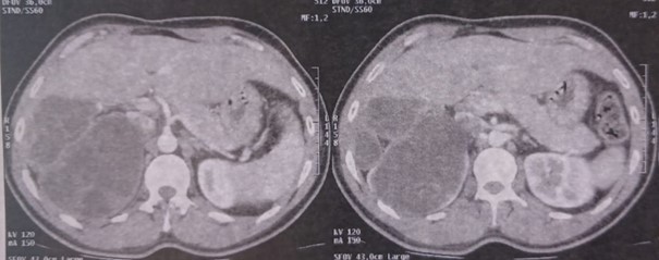
Abdominal CT scan showed a dysmorphic liver with hypotrophy of the left lobe, with a cystic lesion at the site of the left hepatic segmentectomy, well-defined, with regular contours, measuring 88x55x61 mm, and containing multiple septa. Additionally, a large multi-septated cystic formation was observed in the upper pole of the right kidney, measuring 98 x 97 x 100 mm. These cystic formations enhanced after contrast injection and were consistent with Gharbi type III hydatid cysts (Figure 1).
The hydatid serology, performed using the ELISA technique, was positive.
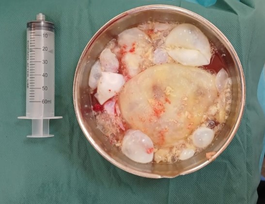
The patient underwent resection of the prominent dome of both the hepatic and renal hydatid cysts, with drainage of the residual cavities (Figure 2).
Post-operative recovery was uneventful. The patient was started on Albendazole 400 mg twice daily and discharged 4 days after the procedure.
On his latest follow-up appointment, 6 months after the surgery, an ultrasound study was requested which showed no evidence of recurrence.

Figure 1: Hepatic and right renal hydatid Cyst.

Figure 2: Evacuation of the proliferative vesicles.
Discussion
Hydatid cysts, caused by the parasitic tapeworm Echinococcus granulosus, represent a major public health concern in Morocco, where the disease remains endemic. The country has a high prevalence of hydatid disease, particularly in rural and agricultural regions, where livestock farming is widespread, and the human-animal interaction is frequent [1,2].
The disease is most commonly found in the liver and lungs, but it can affect other organs such as the brain, bones, and kidneys. Isolated renal hydatid cysts are rare, and 44% of patients with this condition have other concurrent locations. While dual involvement of both the kidneys and liver with hydatid cysts is possible, it remains relatively rare. In most cases, kidney involvement is unilateral, occurring in 85% of instances, as illustrated in this case [3]. Additionally, hydatid cysts are typically more common in the left kidney, unlike the case presented above, where the right kidney was affected [4,5].
Furthermore, imaging is essential for diagnosing and staging hydatid cysts. Ultrasound is considered the most important diagnostic tool for hydatid disease [6]. However, since Morocco is an endemic country with a high risk of recurrence, we opted for a conservative treatment approach in our case.
Antihelminthic treatment alone is not the ideal treatment for hydatid cysts, radical surgery with administration of albendazole is the best treatment option due to low recurrence and complication rates [7].
Despite all efforts to to combat the hydatid disease, the control of hydatid cysts remains a complex challenge due to the rural nature of the affected areas, limited healthcare access in remote regions, and the deep-rooted cultural and economic factors surrounding animal husbandry practices [8]. Therefore, continuous and coordinated efforts are essential to further reduce the burden of hydatid disease in Morocco.
Conclusion
The dual involvement of lung and kidney underscores the importance of considering hydatid disease in endemic regions, even when multiple organ systems are affected. Early diagnosis through imaging techniques, particularly ultrasound, plays a pivotal role in the timely management of such cases. This report also emphasizes the need for increased awareness and preventive measures, including regular deworming of dogs and public education on the risks associated with hydatid disease, to reduce the burden of this parasitic infection in endemic areas like Morocco.
References
- Azlaf R, Dakkak A. Epidemiological study of the cystic echinococcosis in Morocco. Vet Parasitol, 2006; 137(1-2): 83-93. doi: 10.1016/j.vetpar.2006.01.003.
- Brik K, Hassouni T, Youssir S, Baroud S, Elkharrim K, Belghyti D. Epidemiological study of Echinococcus granulosusin sheep in the Gharb plain (North-West of Morocco). J Parasit Dis, 2018; 42(4): 505-510. doi: 10.1007/s12639-018-1026-7.
- Demir M, Yağmur İ. Isolated renal hydatid cyst in a 6-year-old boy: a case report. J. Parasitol, 2021; 16(4): 692–696. doi: 10.18502/ijpa.v16i4.7883.
- Imani F, Gillet J, Benchekroun A, Benomar M, Moreau JF. Aspects radiologiques des kystes hydatiques du rein. A propos de 10 cas vérifiés [Radiological appearances of hydatid cysts of the kidney. 10 confirmed cases (author's transl)] J. Radiol. Electrol. Med. Nucl, 1977; 58(2): 135–144.
- Göğüş C, Safak M, Baltaci S, Türkölmez K. Isolated renal hydatidosis: experience with 20 cases. J. Urol, 2003; 169(1): 186–189. doi: 10.1016/S0022-5347(05)64064-5.
- Stojkovic M, Rosenberger K, Kauczor HU, Junghanss T, Hosch W. Diagnosing and staging of cystic echinococcosis: how do CT and MRI perform in comparison to ultrasound? PLoS Neglected Trop. Dis, 2012; 6(10). doi: 10.1371/journal.pntd.0001880.
- Gomez I Gavara C, López-Andújar R, Belda Ibáñez T, Ramia Ángel JM, Moya Herraiz Á, Orbis Castellanos F, et al. Review of the treatment of liver hydatid cysts. World J Gastroenterol, 2015; 21(1): 124-131. doi: 10.3748/wjg.v21.i1.124.
- Amarir F, Rhalem A, Sadak A, Raes M, Oukessou M, Saadi A, et al. Control of cystic echinococcosis in the Middle Atlas, Morocco: Field evaluation of the EG95 vaccine in sheep and cesticide treatment in dogs. PLoS Negl Trop Dis, 2021; 15(3): e0009253. doi: 10.1371/journal.pntd.0009253.

