Dermoscopy to the Rescue: It’s Not a Bug, It’s Skin Cancer
Saadia Boughaleb*, Zakia Douhi, Meryem Soughi, Sara Elloudi, Hanane Baybay and Fatima Zahra Mernissi
Dermatology and Venereology Department, Hassan II University hospital, Fez, Morocco
Received Date: 07/03/2025; Published Date: 12/05/2025
*Corresponding author: Saadia Boughaleb, Dermatology and Venereology Department, Hassan II University hospital, Fez, Morocco
ORCID iD : 0009-0002-2656-443X
Abstract
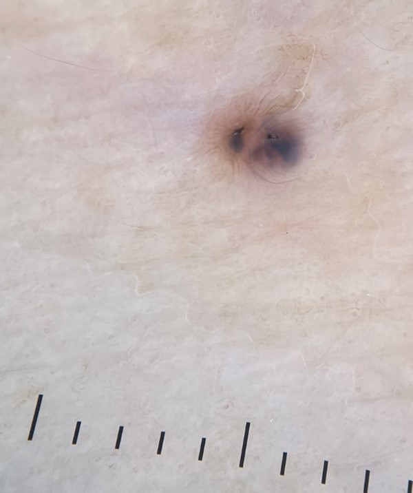
We report the case of an 81-year-old woman with phototype IIIa, presenting with a 3 mm pigmented, non-ulcerated papule was discovered on her inner forearm—an area not typically sun-exposed. Dermoscopy revealed pigmented ovoid nests, globules, micro-erosions on an erythematous background, and peripheral pigmented lines, forming a striking pattern reminiscent of a ladybug. Surgical excision followed by histopathological analysis confirmed a pigmented nodular BCC. This case illustrates several critical teaching points: small basal cell carcinomas can mimic benign lesions and may appear in non-sun-exposed areas; dermoscopy plays an essential role in early recognition and should be integrated into routine full-body skin assessments in high-risk patients. Finally, the diagnostic value of dermoscopy in identifying key features—such as ovoid nests and blue-gray dots—which are strongly correlated with the nodular histologic subtype, especially in lesions under 5 mm.
Keywords: Basal cell carcinoma; Dermoscopy; Skin imaging
Main Text
We report the case of an 81-year-old woman with a phototype IIIa and no significant history of chronic sun exposure. She had a history of multiple basal cell carcinomas (BCCs) on the face, all surgically treated. The patient was regularly followed, with no signs of recurrence at surgical sites and no suspicious lesions in photo-exposed areas.
Given her history of skin cancer, she was educated on self-examination to detect new lesions between consultations. However, she denied noticing any changes. Despite this, a routine full-body examination revealed a 3 mm, non-ulcerated, pigmented papule on the inner forearm, unnoticed by both the patient and her family (Figure 1).
Dermoscopy showed pigmented ovoid nests and globules, micro-erosions on an erythematous background, and peripheral pigmented lines, forming a pattern strikingly similar to a ladybug (Figure 2). The lesion was surgically excised, and histopathology confirmed the diagnosis of pigmented nodular basal cell carcinoma.
Teaching point:
This case highlights the importance of full-body skin examinations rather than limiting assessments to sun-exposed areas, particularly in patients at increased risk for skin cancer. While BCCs are predominantly found on the head and neck due to chronic sun exposure, unusual localizations have been documented in the literature [1]. Our observation reinforces that BCCs can develop in unexpected non-sun-exposed areas.
Furthermore, this case underscores the value of dermoscopy in the early detection of BCCs, even in millimetric lesions that may be overlooked during routine examination. Dermoscopy plays a crucial role in identifying specific patterns, such as pigmented ovoid nests and micro-erosions, allowing for prompt diagnosis and treatment. Studies have shown that small BCCs are histologically correlated with the nodular subtype [2]. In dermoscopy, blue-gray dots and ovoid nests are the most predictive features for diagnosing small BCCs (<5 mm), rather than leaf-like structures or shiny white features [3]. The dermoscopic and histologic findings in our patient align with the literature, and the unique resemblance of the lesion to a ladybug serves as a reminder that BCCs can present with diverse clinical appearances, emphasizing the need for dermoscopic evaluation in all suspicious lesions.
Additionally, this case illustrates the limitations of self-examination. Despite patient education and awareness, small or atypically located lesions may go unnoticed by both patients and their families. This highlights the indispensable role of regular dermatologic follow-ups and systematic skin assessments in high-risk individuals.
Ultimately, our findings reaffirm the need for a comprehensive approach to skin cancer screening, incorporating full-body examinations, dermoscopic evaluation, and patient education to ensure early detection and optimal management of BCCs, regardless of their location or clinical presentation.
Conflict of interest: None
Funding sources: None

Figure 1: Clinical image showing a 3 mm pigmented, non-ulcerated papule on the inner forearm.

Figure 2: Dermoscopic image of the lesion displaying pigmented ovoid nests, globules, micro-erosions on an erythematous background, and peripheral pigmented lines. Notably, the pattern resembles a ladybug.
References
- Schwartzberg L, Arora N. Hidden Basal Cell Carcinoma in the Intergluteal Crease. Cutis, 2021; 107(2): 95-96. doi: 10.12788/cutis.0169.
- Longo C, Specchio F, Ribero S, et al. Dermoscopy of small-size basal cell carcinoma: a case-control study. J Eur Acad Dermatol Venereol JEADV, 2017; 31(6): e273-e274. doi: 10.1111/jdv.13988.
- Arias-Rodriguez C, Muñoz-Monsalve AM, Cuesta D, Mejia-Mesa S, Aluma-Tenorio MS. Dermoscopy of very small basal cell carcinoma (≤3mm). An Bras Dermatol, 2023; 98(6): 755-763. doi: 10.1016/j.abd.2022.12.004

