An Interesting Rare Case of Meckel’s Diverticulum Hernia “Littre’s Hernia”
Majd Abdessamad, Kamal Benzidane*, Soukaina Khafif, Abdelhak Ettaoussi, Kamal Khadija, Mounir Bouali, Abdellillah El Bakouri and Khalid El Hattabi
Hassan II University of Casablanca, Morocco Institution: Department of Visceral Surgical Emergency, Ibn Rochd University Hospital, Casablanca, Morocco
Received Date: 08/02/2025; Published Date: 18/03/2025
*Corresponding author: Kamal Benzidane, Hassan II University of Casablanca, Morocco, Institution: Department of Visceral Surgical, Ibn Rochd University Hospital, Casablanca, Morocco
Abstract
Meckel’s Diverticulum (MD) is a congenital anomaly resulting from incomplete obliteration of the omphalomesenteric duct. While most cases are asymptomatic, complications such as bleeding, bowel obstruction, ulceration, diverticulitis, and perforation can occur. Littre’s hernia refers to the rare protrusion of a Meckel’s diverticulum through a hernial orifice, most commonly in the inguinal, femoral, or umbilical regions. Diagnosis is typically made intraoperatively due to its nonspecific clinical presentation. Surgical management includes laparotomy or laparoscopy, with diverticulectomy or segmental resection performed in complicated cases.
Keywords: Meckel’s diverticulum; Littre’s hernia; Bowel obstruction; Diverticulectomy; Segmental resection; Hernial defect; Surgical management
Introduction
Meckel’s Diverticulum (MD) is a remnant of the proximal portion of the omphalomesenteric duct, which connects the embryonic intestine to the umbilical bladder until the fifth week of gestation. It was first discrebed in the 18th-century by the French anatomist, Alexis de Littre as an ileal diverticulum [1,2].
MD is one of the most common congenital anomalies of the gastrointestinal tract, occurring in approximately 2% of the adult population [3]. It has a complication rate of 4-6%, with potential issues such as gastrointestinal bleeding, bowel obstruction, inflammation, and perforation [4].
The presence of MD in the hernia sac is a rare condition known as Littre’s hernia, with an unknown frequency [5]. Diagnosis is generally made intraoperatively due to the symptomatology, wich is similar to that of other hernia containing small intestine [6,7].
We present the case of a 37-year-old patient, admitted to the emergency room for a strangulated umbilical hernia with an intraoperative diagnosis of Littre’s hernia.
Case Report
A 37-year-old female patient with no significant medical history was admitted to the emergency department for a painful umbilical swelling, accompanied by bilious vomiting, with no other associated digestive symptoms.
On clinical examination, the patient was conscious, hemodynamically and respiratory stable. Abdominal examination revealed a strangulated umbilical hernia with local inflammatory signs. The other hernial orifices were free.
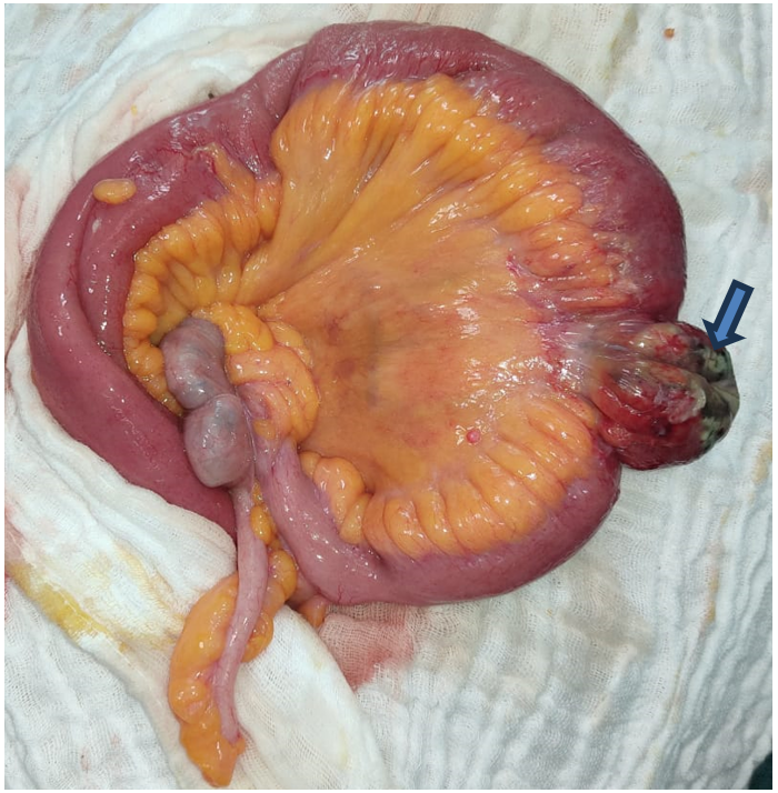
The patient was admitted to the operating room and underwent laparotomy via an arciform incision around the umbilicus. Upon exploration and after opening of the hernial sac, a hernia with a 3 cm neck was found, containing a necrotic Meckel’s diverticulum.

Figure 1: Per operative image showing a necrotic Meckel’s diverticulum.
The surgical procedure consisted of a segmental ileal resection, including the diverticulum, followed by a termino-terminal ileo-ileal anastomosis.
The postoperative was uneventful, with the resumption of bowel function on postoperative day 2 in the form of gas and on day 3 in the form of stool. The patient was discharged on postoperative day 4.
Histopathological examination of the resected specimen revealed nonspecific edematous-congestive lesions.
Discussion
A Meckel’s diverticula are the result of incomplete obliteration of the embryologic omphalomesenteric duct. It is often described by the “rule of 2’s”; occurs in 2% of the population, typically presents before 2 years of age, is approximately 2 inches in length, and it is located within 2 feet of the ileocecal valve. It contains all the normal layers of the intestinal wall, but in 50% of cases, it has some evidence of ectopic gastric, pancreatic, duodenal, colonic, or biliary mucosa [8].
Most cases of MD are clinically asymptomatic, with only 4% to 6% of cases becoming symptomatic. The most common presentation is bleeding due to ectopic gastric mucosa, which occurs more frequently during infancy. In adults, complications such as bowel obstruction (due intussusception or adhesions), ulceration, diverticulitis, and perforation are more common [9-11].
Littre hernia, is the protrusion of a Meckel’s diverticulum through a hernial orifice. It’s a rare condition with an unknown true incidence, but it is estimated to occur in 1% of patients with a known Meckel’s diverticulum [12,13]. The most common localisation of LH is the inguinal region with 50%, followed by the femoral region in 20% and the umbilical region in 20% [14,15].
Two types of LH have been described depending on the contents of the hernia sac. The first type, known as “True LH”, containing only a MD, whereas the second type, “Mixed LH”, includes a MD and other intra-abdominal organs such as the small bowel [4].
the symptomatology of LH does not differ from that of other abdominal wall hernias, which explains why imaging is often unnecessary in this case. As reselt, the diagnosis is most often made intraoperative.
As with other hernias, the surgical approach can be either open (laparotomy) or minimally invasive (laparoscopy).
For complicated cases of MD, resection is the main treatment, performed either as a diverticulectomy or a segmental bowel resection. However, in patients with LH and an asymptomatic MD, it can be left intact, and only the hernial defect can be repaired. Certain indications suggest resection if found incidentally, including patients younger than 40 years of age, the presence of a fibrous band attached to the MD, the presence of heterotopic mucosa, or a long MD with a narrow base. If malignant tumors are found within the MD, a carcinologic bowel and mesentery resection is required [16].
Regarding hernial defect repair, mesh is generally not used in cases of contamination, such as in the presence of incarceration or perforation [4].
Conclusion
Meckel’s diverticulum is a common congenital anomaly, often asymptomatic but capable of causing significant complications. Littre’s hernia, the rare protrusion of a Meckel’s diverticulum through a hernial orifice, remains a challenging diagnosis, typically made intraoperatively due to its nonspecific clinical presentation. Surgical intervention is the mainstay of treatment, with resection reserved for complicated cases or when risk factors are present. Proper management, including appropriate surgical techniques and hernial defect repair, is essential to prevent complications and ensure favorable outcomes.
References
- Opitz JM, Schultka R, Gobbel L. Meckel on developmental pathology. Am J Med Genet Part A, 2006; 140(2): 115-128.
- Kanazawa K, Ishikawa K, Shoji R, Okamoto A. Littre’s femoral hernia causing intestinal fistula. Jpn J Surg, 1972; 2(1): 37-46.
- Matsagas MI, Fatouros M, Koulouras B, Giannoukas AD. Incidence, complications, and management of Meckel’s diverticulum. Arch Surg, 1995; 130(2): 143-146.
- Schizas D, Katsaros I, Tsapralis D, et al. Littre's hernia: a systematic review of the literature. Hernia. 2019, 23:125-30. 10.1007/s10029-018-1867-0.
- Misiak P, Piskorz Ł, Kutwin L, Jabłoński S, Kordiak J, Brocki M. Strangulation of a Meckel’s diverticulum in a femoral hernia (Littre’s hernia). Prz Gastroenterol, 2014; 9(3): 172-174.
- Yagmur Y, Akbulut S, Can MA. Gastrointestinal perforation due to incarcerated Meckel’s diverticulum in right femoral canal. World J Clin Cases, 2014; 2(6): 232-234.
- Park JJ, Wolff BG, Tollefson MK, Walsh EE, Larson DR. Meckel diverticulum: the Mayo Clinic experience with 1476 patients (1950-2002). Ann Surg, 2005; 241(3): 529-533.
- Skandalakis PN, Zoras O, Skandalakis JE, Mirilas P. Littre hernia: Surgical anatomy, embryology, and technique of repair. Am Surg, 2006; 72: 238-243.
- Sagar J, Kumar V, Shah DK. Meckel’s diverticulum: A systematic review. J R Soc Med, 2006; 99: 501-505.
- Hansen C-C, Søreide K. Systematic review of epidemiology, presentation, and management of Meckel’s diverticulum in the 21st century. Medicine (Baltim), 2018; 97: e12154.
- Moore TC. Omhalomesenteric duct malformations. Semin Pediatr Surg, 1996; 5: 116-123.
- Usman A, Rashid MH, Ghaffar U, Farooque U, Shabbir A. Littré’s hernia: A rare intraoperative finding. Cureus, 2020; 12: e11065.
- Schizas D, Katsaros I, Tsapralis D, Moris D, Michalinos A, Tsilimigras DI, et al. Littre’s hernia: A systematic review of the literature. Hernia, 2019; 23: 125-130.
- ô. Yagmur Y, Akbulut S, Can MA. Gastrointestinal perforation due to incarcerated Meckel's diverticulum in right femoral canal. World J Clin Cases, 2014; 2(6): 232-234. doi:10.12998/wjcc.v2.i6.232.
- Castleden WM. Meckel's diverticulum in an umbilical hernia. Br J Surg, 1970; 57(12): 932-934. doi:10.1002/bjs.1800571216.
- Evola G, Piazzese E, Bonanno S, Di Stefano C, Di Fede GF, Piazza L. Complicated Littre's umbilical hernia with normal Meckel's diverticulum: a case report and review of the literature. Int J Surg Case Rep, 2021; 84: 106126. doi: 10.1016/j.ijscr.2021.106126.

