Unilateral Congenital Venous Malformation in the Form of Giant Right Saphena Varix and Proximal Varicose Veins in a Toddler Boy
Govani ND1, Govani DR1, Panchasara NG1 , Patel RR1,*, Midha PK2 and Patel RV1
1Department of Pediatrics and Pediatric Surgery, Postgraduate Institute of Child Health & Research and KT Children Govt University Teaching Hospital, Rajkot 360001, Gujarat, India
2J. Watumull Global Hospital & Research Centre, Delwara Road, Mount Abu, Rajasthan 307501, India affiliated to medical faculty of God fatherly spiritual university, Mount Abu, Rajasthan, India
Received Date: 16/10/2024; Published Date: 08/11/2024
*Corresponding author: Mr. Ramnik Patel MD, MS, MCh, LL M, DCH, DNBPS, FRCSEd, FRCS Ped, FEBPS, FACS, FAAP, Director-Professor, Department of Pediatric Surgery, Postgraduate Institute of Child Health and Research and K T Children Government University Teaching Hospital Rajkot 360005, Gujarat, India
Abstract
An interesting rare case of unilateral congenital venous malformation in the form of giant right sided saphena varix and proximal varicose veins in a 2½-year-old boy has been reported. The patient presented with a right groin mass and varicosities of the proximal great saphenous vein. Imaging studies by ultrasound and color doppler scan confirmed the findings. Gradual increasing of the mass, pain and cosmetical considerations were indications for surgery in the form of high ligation at sapheno-femoral junction, triple ligation of the main tributaries and excision of the giant right saphena varix and interventional procedures like sclerotherapy and endo-laser for the right proximal saphenous varicosities with excellent results.
Keywords: Congenital; Endo-laser; Saphena varix; Sclerotherapy; Triple ligation; Varicose veins; Venous malformations; Vascular anomalies
Introduction
Congenital vascular anomalies are a broad-spectrum term which includes two of the most common congenital vascular anomalies, namely, hemangiomas and vascular malformations which superficially and externally may look similar but have several differences between both of these lesions [1]. Congenital vascular malformations have arterial, venous, capillary and mixed varieties and congenial isolated venous malformation in the form of saphena varix and varicosities are very unusual and exceptional unilaterally in a toddler [2-4]. Varicose veins are common in elderly, females; affect the distal long saphenous vein initially, can result in significant circulatory disease and are extremely rare in children [5-9]. A case of a varicose malformation of the saphenofemoral junction and proximal long saphenous vein in a 2½-year- old boy is hereby presented. To the best of our knowledge, no such case was previously
reported in literature and use of advanced technology like endo-laser and ultrasound guided sclerotherapy has successfully been used with excellent cosmetic and functional results.
Case Report
A 2½-year-old boy was referred to the pediatric surgery clinic by the plastic surgical team with a chief complaint of a mass in the right groin and vascular blemish on his right thigh present since birth. He does occasionally say that it is sore, but otherwise he appeared relatively asymptomatic. Subsequently this has progressed up the thigh terminating in a lump in his groin which was seen especially during times of crying or exertion. The mass gradually increased in size. The child was born after normal pregnancy and delivery. The parents ruled out any previous trauma. The patient has mild asthma and mother’s grandmother has mild varicose veins developed at the age of 60 treated conservatively.
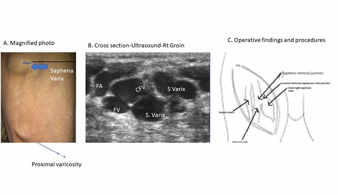
On examination, a 3×3 cm2 compressible mass with a bluish tinge was noticed in the right groin. The mass increased in size on cough impulse and disappeared on lying down. On auscultation a venous hum was heard. The skin over the lateral aspect of
the right thigh was covered by multiple superficial varicosities in different sizes progressing down and stopping at the knee in the line of the long saphenous vein (Figure 1A).
Ultrasound and venous doppler mapping confirmed the clinical findings. It showed gross incompetence at the right saphenofemoral junction and varicosities above knee from perforators and normal deep venous system (Figure 1B). Right side below knee and left side superficial and deep venous systems were completely normal. His case was discussed at the vascular anomaly’s clinic in detail.
The patient underwent initial endovenous laser therapy (EVLT) with partial response.
Ultrasound guided access using 4F sheath was used. Total of 8 Cms of anomalous vein was treated stopping 2 cms before the junction with femoral vein and total of 430 joules (10 watts) were delivered. Post therapy, saphena varix was identifiable when he stood upright and at which point a large sac filled in the right groin very slowly with a turbulent flow from a very narrow neck onto the saphenofemoral junction. Unlike previously, the varix did not appear to tract down the thigh at all and there was discoloration to the lateral aspect above the right knee with a palpable cord suggesting a thrombosis in the obliterated superficial varicose veins.
Subsequently patient underwent high ligation of sapheno-femoral junction with flush triple ligation with flush ligation of all three main tributaries and excision of the giant saphena varix and ultrasound guided injection sclerotherapy. At surgery the groin was explored by a transverse incision (Figure 1C). A large saphena varix with all three dilated and very tortuous tributaries were found. The medial tributary was normal. There was plenty of perivenous and peri varix fibrosis. Sclerotherapy of the superficial varicosities in the distal right thigh was carried out under ultrasound guidance using total of 1 ml of 0.5% sodium tetradecylsulphate. The postoperative course was unremarkable.
At follow up, patient had pleasing results with disappearance of the saphena varix and the varicosities of veins. He had some superficial pigmentation over the skin in the lateral aspect of the right thigh following local sclerotherapy. Ultrasound and Doppler studies showed post operative changes with resolution of the saphena varix and varicosities.

Figure 1A: Clinical Photograph-Right Saphena Varix and Varices over the right thigh. 1B: Cross sectional ultrasound scan demonstrating saphena varix and incompetent saphenofemoral junction. With giant saphena varix- FA-Femoral artery, FV-Femoral vein, CFV- Common femoral vein, S Varix-Saphena varix. 1C: Operative findings and procedure- High ligation at saphenofemoral junction with triple ligation and excision of giant saphena varix.
Discussion
Varicose veins are abnormally dilated, elongated and tortuous, veins. There are various causes of varicose veins in the lower extremities. There are primary and secondary causes of varicose veins. Primary causes are congenital and/or develop from inherited conditions. Secondary
causes generally result from factors other than congenital factors. Congenital factors include incomplete or absence of valves, and incompetent fibrous or elastic tissues in the vein wall [10]. Incompetent fibrous or elastic tissues in the vein wall are congenital and developmental.
The role of heredity in the development of varicose veins of the lower limbs has been raised many times in the literature. To the best of our knowledge, no previously reported case of congenital saphena varix and proximal saphenous varicose veins has been documented. The risk of developing varicose veins for the children was 90% when both parents suffered from this disease, 25% for males and 62% for females when one parent was affected, and 20% when neither parent was affected [11]. Multiple pregnancies, old age, and white race are known risk factors. Regardless of the epidemiologic and pathophysiologic features, the pathogenesis of varicose veins in the lower extremities is mainly venous valvular dysfunction.
A saphena varix, or a saphenous varix is a dilation of the saphenous vein at its junction with the femoral vein in the groin. As saphena varix and varicose is usually asymptomatic or have mild symptoms, the indications for surgery are discomfort and progressive expansion involving cosmetic considerations, as was the case in our patient. Varicosities are generally seen in the lower limbs but unusual locations include vulvoperineal varicosity, intraosseous perforating vein incompetence, round ligament varicosity, persistent sciatic vein incompetence, Klippel-Trenaunay syndrome, and portosystemic collateral pathways.
Traditionally, Doppler ultrasonography (US) has been used for evaluation of varicose veins. Sometimes, varicose veins arise from an unexpected anatomic source; in these cases, computed tomographic (CT) venography can provide an overview of the varicose veins. Doppler US with complementary CT venography is useful for determining the precise cause of varicose veins.
Radiologists should be familiar with the complete range of primary causes of varicose veins in the lower extremities and with their radiologic manifestations and should recognize the complementary role of CT venography in their evaluation.
Endovenous laser treatment is a recent advance in the treatment of varicose veins. Severe cases may be combined with ultrasound guided sclerotherapy and surgical ligation [12]. The results are excellent and the prognosis is very good.
Conclusion
In conclusion, our case is an unusual and a rare presentation of a congenital vascular anomaly in the form of giant right sided saphena varix and right proximal saphenous varicosities which was diagnosed promptly in the immediate post-natal period using ultrasound and color doppler and treated successfully by combination of various interventional procedures like endo-laser and sclerotherapy for the varicose veins and surgical high flush ligation at the sapheno-femoral junction with triple ligation of the three main tributaries and excision of the giant saphena varix with excellent cosmetic and functional results and very good long term prognosis.
References
- Patel RV, Curry JI, Sinha CK, Davenport M. Vascular Anomalies. In: Sinha, C.K., Davenport, M. (eds) Handbook of Pediatric Surgery. Springer, Cham, 2022. https://doi.org/10.1007/978-3-030-84467-7_62 Springer Verlag, London, 2021; pp 523–529. https://link.springer.com/chapter/10.1007/978-3-030-84467-7_62.
- Patel R, Curry JI, Sinha CK, Davenport M. Vascular Malformations. In: Handbook of Pediatric Surgery. Part 4 Chap 13, 1st Ed, CK Sinha & Mark Davenport (Eds), Springer Verlag, London, 2010, 221-228. ISBN-10. 184882131X · ISBN-13. 978- 1848821316 · Edition. 2010th · Publisher. Springer, 2010.
- Govani DJ, Govani ND, Govani DR, Panchasara NG, Patel RR, Midha PK, et al. Congenital giant progressive parotid hemangioma in an infant treated successfully with propranolol and steroids. J Clin Images Med Case Rep, 2023; 4(3):
- Patel RV, Govani DJ, Govani ND, Govani DR, Panchasara NG, Patel RR, et al. Giant cavernous vascular venous malformation of glans and distal penile shaft in a toddler treated successfully with steroids followed by circumcision. MedP J Cardiology and Vascular Medicine.2022, Manuscript ID-MPCVM-202205003.
- Samuel M, Spitz L. Klippel-Trenaunay syndrome: clinical features, complications and management in children. Br J Surg, 1995; 82(6): 757-761.
- Steven M, Kumaran N, Carachi R, Desai A, Bennet G. Hemangiomas and vascular malformations of the limb in children. Pediatr Surg Int, 2007; 23(6): 565-569.
- Griton P, Schadeck M. Hyperplasia of the saphenous vein and the initial signs of varicose disease in children. Phlebologie, 1990; 43(4): 561-571.
- Weindorf N, Schultz-Ehrenburg U. The development of varicose veins in children and adolescents Phlebologie, 1990; 43(4): 573-577.
- Griton P. The 1st clinical signs of venous insufficiency in children. Phlebologie, 1992; 45(4): 501-507.
- Ersoy U, Güvener M, Kaplan S, Ozkaya O, Uzunalimoğlu B. Hitherto. undescribed venous vacuolar myopathy without mucoid degeneration in the varicose saphenous vein of a child. Vasa, 2001; 30(1): 67-70.
- Cornu-Thenard A, Boivin P, Baud JM, De Vincenzi I, Carpentier PH. Importance of the familial factor in varicose disease. Clinical study of 134 families. J Dermatol Surg Oncol, 1994; 20(5): 318-326.
- Huang Y, Jiang M, Li W, Lu X, Huang X, Lu M. Endovenous laser treatment combined with a surgical strategy for treatment of venous insufficiency in lower extremity: a report of 208 cases. J Vasc Surg, 2005; 42(3): 494-501.

