Eye of the Tiger Sign
Chaimae Lahlou*, Ihssan Hadj Hsain, Chaymae Faraj, Sara Essetti, Nazik Allali, Siham El Haddad and Latifa Chat
Pediatric Radiology Department, Children’s Hospital, Faculty of Medicine and Pharmacy, Mohammed V University, Rabat, Morocco
Received Date: 01/10/2024; Published Date: 07/11/2024
*Corresponding author: Chaimae Lahlou, Pediatric Radiology Department, Children’s Hospital, Faculty of Medicine and Pharmacy, Mohammed V University, Rabat, Morocco
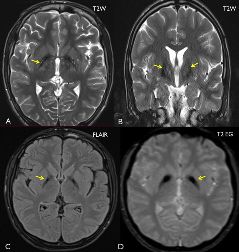
An "eye-of-the-tiger" sign is a distinctive pattern observed in magnetic resonance imaging (MRI), serving as a critical diagnostic indicator of pantothenate kinase-associated neurodegeneration (PKAN). This sign manifests as low-signal intensity rings encircling central regions of high-signal intensity within the medial aspect of the bilateral globus pallidus on T2-weighted MRI scans (Figure 1). The diminished signal intensity surrounding the globus pallidus is attributed to the accumulation of excess iron. The central areas of heightened signal intensity are likely a result of gliosis. PKAN, previously referred to as Hallervorden-Spatz syndrome, is among the three extrapyramidal disorders linked to an elevated presence of brain iron, collectively known as neurodegeneration with brain iron accumulation (NBIA).
There are no formalized criteria for the sign and other conditions, such as Wilson disease, atypical parkinsonism, and organophosphate poisoning may demonstrate a similar appearance. It may also be a normal finding on 3 T MRI. Therefore, caution should be used when interpreting this sign.

Figure: Transverse T2-weighted (A) , Flair(C), T2 EG (D) and coronal T2-weighted (B) MR images showing a low signal intensity ring surrounding a central high intensity region in the globus palludis, producing an eye of the tiger sign.
References
- Chang CL, Lin CM. Eye-of-the-Tiger sign is not Pathognomonic of Pantothenate Kinase-Associated Neurodegeneration in Adult Cases. Brain Behav, 2011; 1(1): 55-56. doi: 10.1002/brb3.8.
- Litwin T, Karlinski M, Skowrońska M, Dziezyc K, Gołębiowski M, Członkowska A. MR image mimicking the "eye of the tiger" sign in Wilson's disease. J Neurol, 2014; 261(5): 1025-1027. doi: 10.1007/s00415-014-7322-y.

