Cesarean Scar Endometriosis
Sara Ez-zaky*, Khaoula Boumeriem, Ihssan Hadj Hsain, Nazik Allali, Latifa Chat and Siham El Haddad
Department of Radiology Mother and child, the children’s hospital, Morocco
Received Date: 04/07/2024; Published Date: 28/10/2024
*Corresponding author: Sara Ez-zaky, Department of Radiology Mother and child, the children’s hospital, Morocco
Clinical Medical Image
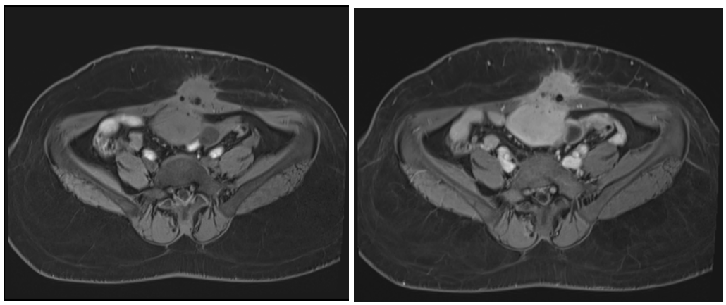
She is a 36-year-old woman, G1P1E1 with a history of cesarean section delivery, presenting with cyclic pelvic pain at the site of the scar. On clinical examination, a mass is palpable at the site of the cesarean scar. An ultrasound was performed, revealing a well-defined heterogeneous mass containing hyperechoic spots. Subsequently, a pelvic MRI was conducted, showing a mass on the anterior abdominal wall, lateral to the left of the umbilicus, oval-shaped with irregular borders. It exhibited heterogeneous intermediate signal intensity on both T1 and T2 sequences, containing small microcystic formations and calcifications with low signal intensity. The mass enhanced after contrast injection, showed no diffusion restriction, and measured 31x38x30mm ,It infiltrates the rectus abdominis muscle of the left abdomen, appears adhered to the uterine fundus with loss of separation margin, and also shows adhesions with low T2 signal in the fat (Figure 1, 2).

Figure 1: Axial and coronal T2-weighted sequences showing: a mass in the anterior abdominal wall, sub-umbilical and lateralized to the left, oval-shaped with irregular contours, heterogeneous intermediate T2 signal, containing small microcystic formations.

Figure 2: Axial T1-weighted sequence before and after gadolinium injection showing: the presence of a mass in the anterior abdominal wall, sub-umbilical and lateralized to the left, oval-shaped with irregular contours, heterogeneous intermediate T1 signal, and calcifications with low signal intensity. The mass enhanced after contrast injection and infiltrates the left rectus abdominis muscle, appearing to be adherent to the uterine fundus.
Scar endometriosis, also referred to as incisional endometrioma, is an uncommon type of extra-pelvic endometriosis that develops within surgical scars where endometrial tissue may become implanted [1]. Cesarean section scars represent the most frequently affected site for scar endometriosis. [2].
Ultrasound is less sensitive than MRI for the evaluation of deep pelvic endometriosis. MRI played a crucial role in diagnosing endometriosis in this case by revealing specific signal characteristics across different sequences, indicating the presence of blood products within the uterine lesion. Additionally, diagnostic findings suggestive of adenomyosis were also observed. Interestingly, preoperative consideration did not include the involvement of the lower segment uterine Cesarean scar. However, histopathological examination confirmed the presence of endometriosis at the Cesarean scar site [3].
References
- Tangri MK, Lele P, Bal H, Tewari R, Majhi D. Endométriose cicatricielle : une série de 3 cas. J. Armed Forces India, 2016 ; 72 (Suppl 1): S185. Disponible sur: pmc/articles/PMC5192233/.
- Jabri K. Endométriose sur cicatrice de césarienne. Med. J,2009; 24(4): 294. Disponible sur: /pmc/articles/PMC3243870/.
- Ashim K Lahiri, Kiran Sharma, Naser Busiri. Endometriosis of the uterine cesarean section scar: A case report, Indian J Radiol Imaging, 2008; 18(1): 66–68. doi: 4103/0971-3026.37111.

