Acute Intussusception on a Lipoma of the Left Colon in an Adult
Rabileh Madina*, Sidki Kenza, Lanjerie Safae, Insumbo Paulino, Wen-Yam Traoré, Saleheddine Tariq, Zamani Ouijdane, Edderai Meryem and El Fenni Jamal
Department of Radiology, Mohammed V of the Military Hospital, Rabat, Morocco
Received Date: 14/08/2024; Published Date: 23/10/2024
*Corresponding author: Rabileh Madina, Department of Radiology, Mohammed V of the Military Hospital, Rabat, Morocco
Abstract
Acute colonic intussusception in adults is rare, accounting for around 5% of intestinal obstructions in adults according to the literature. The causes are organic and intussusception on a lipoma is exceptional. We report a rare case of acute intestinal intussusception caused by a submucosal lipoma of the left colon in a 58-year-old man, with the diagnosis suggested by ultrasound and confirmed by abdominopelvic CT scan. Diagnosis was confirmed during operative reduction and resection. He had segmental left colectomy. The postoperative course was uneventful.
Keywords: Colonic lipoma; Intussusception; CT scan; Treatment
Introduction
Acute Intestinal Intussusception (AII), or even colocolic intussusception in adults, is rare and often secondary to an organic lesion. It accounts for less than 5% of acute intestinal obstructions in adults [1]. In contrast, in children, it represents more than 90% [1,2].
The colonic lipoma, a benign tumor, is rarely encountered in routine clinical practice. Its colonic location is uncommon, accounting for only about 4% of benign tumors of the colon according to the literature [1,4], with its location more frequently being ileal and less often colonic. Usually asymptomatic, colonic lipoma rarely leads to intestinal obstruction caused by colo-colic intussusception. Endoscopic ultrasound, computed tomography, or MRI assist in the diagnosis, which is then confirmed by histology [1-4].
Observation
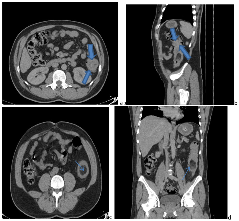
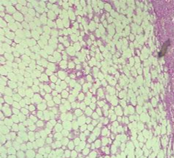
A man aged 58 years with a history of hypertension and Appendicectomy 2 years ago, admitted to the emergency room for epigastric abdominal pain with persistent colicky type pain that is not relieved by analgesics, without any history of vomiting or cessation of bowel movements and gas. On clinical examination, the patient is afebrile, in good general condition, and hemodynamically stable. On examination, the abdomen is diffusely tender, more pronounced in the epigastric region and left upper quadrant, without abdominal rigidity or guarding, and the hernial orifices are free. The laboratory tests did not reveal any biological inflammatory syndrome. We were referred to our department for an abdominal ultrasound, which revealed a target-like image in the left colon containing a hyperechoic mass measuring 25 mm in the largest axis. A follow-up abdominopelvic CT scan with contrast enhancement revealed a target-like image with a central hypodense halo (Figure 1A) and a sandwich-like image topped with a hypodense halo (Figure 1B), well-defined in the upper third of the left colon. This was consistent with a typical image of left colocolic intussusception, showing a well-defined endoluminal mass with homogeneous fat density that did not enhance after contrast injection, measuring 33 mm in its longest axis, adjacent to the intussusception (Figure 1C, 1D). The radiological diagnosis suggested was a left colonic lipoma complicated by colocolic intussusception.Segmental colectomy with colorectal anastomosis was performed . The postoperative course was uncomplicated. Pathology confirmed lipoma without signs of malignancy (Figure 2).

Figure 1: Abdominopelvic CT scans without and with contrast injection showing a target-like image with a central hypodense halo (Figure 1A) and a sandwich-like image topped with a hypodense halo (Figure 1B) with endoluminal mass with homogeneous fat density that did not enhance after contrast injection in axial (1C) and coronal (1D) reconstructions.

Figure 2: Tumoral proliferation of lobules of mature adipocytes without cytonuclear atypia (submucosal lipoma of the colon.
Discussion
Intussusception is more common in children than in adults. In fact, adult intussusception accounts for less than 5% of all cases of intussusception and 1% to 3% of all cases of intestinal obstruction [2]. It affects the small intestine in 48% to 70% of cases, both the ileum and cecum in 25% to 40% of cases, as well as the colon alone in only 5%-8% of cases [5]. Unlike in the child where symptomatic intussusception is almost always idiopathic, in adults symptomatic intussusception is most frequently secondary to an endo-luminal lesion [6].
These lesions are often carcinoma; rarely, it may be benign tumors [3]. Adenomas are the most common colonic benign lesions, while colonic lipomas are less common [1]. They constitute 10% of benign tumors of the digestive tube and 2% to 4% of colon benign tumors. Lipomas can theoretically affect the entire digestive tract, from the hypopharynx to the rectum. In the colon, they are more common in the cecum and ascending colon, and rarer in the left colon. Colonic lipomas primarily affect women aged 50 to 70, are often solitary and submucosal.
They frequently remain asymptomatic and are therefore incidentally discovered during imaging studies, colonoscopies, surgeries, or autopsies. Symptoms, though rare, are more common with pedunculated lipomas larger than 3 cm. These may include abdominal pain, irregular rectal bleeding, anemia, and digestive disorders such as constipation or diarrhea and ultimately leading to an obstructive syndrome. In rare cases, a lipoma can cause acute intussusception [7].
Ultrasonography may be able to diagnose intussusception and lipoma. However, in adults, its performance is limited by the interposition of bowel gas and the corpulence of the patient.
However, it can also show the typical appearance of intestinal intussusception as a "target sign," characterized by two peripheral hypoechoic rings and a central echogenic ring, and in transverse section, as a "sandwich sign," with three superimposed cylinders, which correspond to the intussusceptum and the causative lesion and lipoma is in the form of a regular hyperechogenic lesion surrounded by normal intestinal wall [9]. Abdominal CT is more effective than ultrasound and sensitive and specific for diagnosing intussusception, showing a "bowel within bowel" appearance and associated bowel obstruction. In cases of intussusception caused by lipoma, CT reveals central fat density within the intussusceptum [8].
Regarding histology, a pathological examination is necessary to confirm the diagnosis of lipoma [10,11]. As in our case, analysis of the surgical specimen or biopsies consistently shows the presence of mature adipose cells without any signs of cytonuclear atypia.
In terms of treatment, in adults, the management of intussusception caused by a lipoma is always surgical and does not allow for reduction by radiological control hyperpressure, due to the frequency of underlying organic causes. A more or less extensive resection may be necessary.
Conclusion
Symptomatic intussusception is rare in adults, and colonic lipoma is rarely the cause. In these rare cases, CT is important for diagnosis as it clearly demonstrates the presence and location of intussusception and the fat-density of the lipoma.
References
- Bruère-Ronzi L, Mazet P, Schotte T. Intestinal intussusception in adults. Ann. Fr. Med. Urgence, 2015; 5: 263-264.
- Elhattabi K, Bensardi F, Khaiz D, Fadil A, Raouah A, Lefriyekh R, et al. Intestinal intussusception in adults: a report of 17 cases. Pan Afr Med J, 2012; 12: 17.
- Mnif L, Amouri A, Tahri N. Giant colonic lipomas. Acta Endoscopica, 2010; 41: 1-4.
- Lebeau R, Koffi E, Diané B, Amani A, Kouassi J-C. Acute intestinal intussusception in adults: analysis of 20 cases. Ann Chir, 2006; 131(8): 447-450.
- Abou-Nukta F, Gutweiler J, Khaw J, et al. Giant lipoma causing a colo-colonic intussusception. Am Surg, 2007; 73(4): 417.
- Leon KE, John DC, Arthur HA Jr. Intussusception in Adults: Institutional Review. J Am Coll Surg, 1999; 188: 390-395. http://dx.doi.org/10.1016/S1072-7515(98)00331-7
- Wulff C, Jespersen N. Colo-colonic intussusception caused by lipoma. Acta Radiol, 1995; 36: 478-480.
- Crozier F, Portier F, Wilshire P, et al. CT scan diagnosis of colocolic intussusception caused by a lipoma of the left colon. Ann Chir, 2002; 127: 59-61. http://dx.doi.org/10.1016/S0003-3944(01)00670-8
- Lebeau R, Koffi E, Diané B, et al. Acute intestinal intussusception in adults: analysis of a series of 20 cases. Ann Chir, 2006; 131: 447-450. http://dx.doi.org/10.1016/j.anchir.2006.04.007
- Baba H, Ait Ali A, Damiri A, Elfahsi M, Elhajjouji A, Bouchentouf SM, et al. Sigmoid colon lipoma causing colocolic intussusception. J. Afr. Hepatol. Gastroenterol, 2011; 5: 255-256.
- Goasguen N, Cattan P, Godiris-Petit G, Munoz-Bongrand N, Allez M, Lemann M, et al. Colonic lipoma: a clinical case and literature review. Gastroenterol Clin Biol, 2008; 32: 521-524.

