Pseudo-Aneurysm of the Posterior Tibial Artery: Atypical Localisation of an Artery Pseudoaneurysm
Rachida Chehrastane*, Sanae Jellal, Sara Essetti, Kaouthar Sfar, Fatima Zahra Laamrani and Jroundi Laila
Emergency Radiology Department-Ibn Sina Hospital, Mohammed V University Rabat, Morocco
Received Date: 12/04/2024; Published Date: 03/09/2024
*Corresponding author: Rachida Chehrastane, Emergency Radiology Department-Ibn Sina Hospital, Mohammed V University Rabat, Morocco
Pseudoaneurysms involving infrapopliteal artery are unfrequent. The most common site for lower limb extremity PSA is the anterior artery and rarely the posterior artery [1]. They are usually post-traumatic or iatrogenic [2,3]. Other factors were described, including, arteriosclerosis, Behcet’s disease, hemophilia and osteogenesis imperfecta, type IV of fibromuscular dysplasia, Ehlers-Danlos syndrome, Marfan’s disease.
In addition, other factors such as immunosuppression, malnutrition, and diabetes are also associated with the risk of PSA development [4].
The most common clinical presentation is pain, swelling, paresthesia (which is rare but has been described in the literature), pulsatile or pulseless mass, PSA may be asymptomatic and slightly delayed due to incidental findings during arteriography of know atherosclerotic disease [5,6].
The diagnosis is based on contrast enhanced Computed tomography, which allows to identify the morphology and dimensions, location, the search for complications such as fissure or rupture [2,7].
There is currently no consensus on general treatment strategies for PSA. The choice of treatment is influenced by the size and location of the PSA. It also depends on the patient's medical condition, clinical manifestations of PSA. With spontaneous improvement, smaller asymptomatic PSA values may be observed. However, larger symptomatic masses require resolution, treated endovascularly or surgically [8].

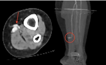
Figure: CT scan of a 65-year-old female patient with a history of hypertension under treatment, diabetes mellitus, and a femoro-femoral bypass surgery following total thrombosis of the left common iliac artery, presented intense pain and paresthesia of the left lower limb.
A: Axial contrasted enhanced Ct image showing a saccular bulge of the posterior left tibial artery
B: 3D reconstruction showing a pseudoaneurysm of the tibial artery, posterior with no enhancement of the left posterior tibial artery beyond the aneurysm and anterior tibial artery due to atheromatous overload.
References
- Miwa T, Ikeda S, Muroya T, et al. Anterior tibial artery rupture treated using covered stent in a patient with vascular Ehlers–Danlos syndrome. J Cardiol Cases, 2018; 18: 197–
- Murphy A, Chan M, Fairbank Tibial nerve palsy as the presenting feature of posterior tibial artery pseudoaneurysm. ANZ J Surg, 2018; 88: 1206–1208.
- Yammine K, Kheir N, Daher J, et Pseudoaneurysm following ankle arthroscopy: a systematic review of case series. Eur J Orthop Surg Traumatol, 2019; 29: 689–696.
- Yu JL, Ho E, Wines Pseudoaneurysms around the foot and ankle: case report and literature review. Foot Ankle Surg, 2013; 19: 194–198.
- van Helden EJ, Eefting D, Florie J, et Endovascular salvage of a false aneurysm of the posterior tibial artery caused by a stab from a stingray. Cardiovasc Intervent Radiol, 2015; 38: 498–500.
- Kalyan JP, Kordzadeh A, Hanif MA, et al. Nonunion of the tibial facture as a consequence of posterior tibial artery J Surg Case Rep, 2015; 2015.
- Haber LL, Thompson G, DiDomenico L, et al. Pseudoaneurysm of the perforating peroneal artery after subtalar joint injury: a case Foot Ankle Int, 2008; 29 :627–629.
- Ellis Inferior medial geniculate artery pseudoaneurysm complicating arthroscopic partial meniscectomy. ANZ J Surg, 2018; 88: E619–E620.

