Management of Mixed Melasma on Darker Phototypes: A New and Safe Laser Therapy Treatment
Massimo DF Vitale*
Private practice, Cosmetic Doctor, Bologna, Italy
Received Date: 21/01/2024; Published Date: 04/06/2024
*Corresponding author: Massimo DF Vitale, MD, Private practice, Cosmetic Doctor, Bologna, Italy
Abstract
Background: Melasma is a skin pigmentation disorder characterized by dark spots and patches on the face and other skin often exposed to the sun. While it can affect all people, people with darker skin have a significantly higher risk of developing this discoloration. In some parts of the world, such Southeast Asia and Latin America, up to 30% of people may suffer from melasma, an acquired skin condition. In fact, individuals with darker Fitzpatrick phototypes IV–VI frequently have hyperpigmented macules on their faces, particularly women (90% of patients). It is characterized by uneven borders and bilateral distribution in sun-exposed areas, especially the face. The three most typical types of melasma are mandibular, malar, and Centro facial.
Materials and Methods: Four patients were treated for facial melasma. Up to two sessions with variable intervals were performed with a 675-nm laser device. The pain during treatment was measured using a Visual Analog Scale of 10 points. The non-ablative laser system used emits red light with a wavelength of 675 nm through a 15 × 15 mm scanning system.
Results: At T1, a consistent improvement in the pigmentary and vascular components was visible. This is always combined with a considerable reduction in vascular expression.
Conclusion: The purpose of this case series is to support the scientific literature on the application of a 675-nm laser device for the treatment of vascular and pigmentary symptoms associated with facial melasma in both men and women. The author believes that the laser can be very effective for benign pigmented lesions, lowering the risk of side effects and simplifying post-treatment management because of its strong affinity for collagen and melanin as well as the typical anatomical capillary structure.
Keywords: Regenerative medicine; 675-nm laser; Facial melasma; Phototypes III to IV (or darker phototypes)
Introduction
Melasma is a skin pigmentation disorder characterized by dark spots and patches on the face and other skin often exposed to the sun. While it can affect all people, people with darker skin have a significantly higher risk of developing this discoloration [1]. In some parts of the world, such Southeast Asia and Latin America, up to 30% of people may suffer from melasma, an acquired skin condition [2]. In fact, individuals with darker Fitzpatrick phototypes IV–VI frequently have hyperpigmented macules on their faces, particularly women (90% of patients). It is characterized by uneven borders and bilateral distribution in sun-exposed areas, especially the face. The three most typical types of melasma are mandibular, malar, and centrofacial [3].
Using Wood's lamp examination, melasma is classified as epidermal, dermal, or mixed form depending on where the pigment is located.While ultraviolet light exposure and hormonal impacts appear to be the most influential variables in the development of symptoms, the exact cause is likely to be complicated and not well understood [4].
Melasma often causes adverse psychological impacts on patients and diminishes their quality of life. Patients often experience feelings of humiliation, low self-esteem, boredom, dissatisfaction, and a lack of social motivation [5-7].
Discussion
Therapeutic management of melasma is challenging, with high recurrence rates which significant impacts on the quality of life. No single treatment is universally efficacious. Systemic treatments with tranexamic acid and polypodium leucotmatous had promising results, although the former was related to systemic side effects. Microneedling and peeling were also efficacious, although their superiority to topical hydroquinone, the gold standard in melasma treatment, remains to be established [8,9].
In contrast to the first-line treatments, laser therapy has recently emerged as a feasible alternative for people who have melasma that has not responded to other treatments. Numerous clinical trials have been conducted up to this point to investigate the efficacy of a wide range of different laser therapies as well as the adverse effects that they may cause. Lasers and light therapy can be broken down into five primary categories: intense pulsed light (IPL), Q-switched lasers, picosecond lasers, non-ablative fractionated resurfacing lasers, and ablative fractionated resurfacing lasers. They have been shown to be quite successful; nevertheless, the downtime generally lasts for a long time, and individuals are unable to instantly return to their regular lives [10].
Previous studies have shown that the 675-nm wavelength is optimal for addressing acne scars, face aging, and skin resurfacing, as stated in the available literature [11]. Its emission in the red spectrum strongly binds to collagen fibers and melanin chromophore, while having minor interaction with the vascular chromophore [12].
By reducing the contact with other chromophores, the laser beam efficiently transmits heat to the collagen fibers, causing them to shrink and undergo denaturation. This process has been histologically confirmed to stimulate the formation of new collagen (neocollagenogenesis) [13].
Bonan and colleagues showed the effectiveness in patients with Fitzpatrick phototypes IV to V, the 675-nm laser system appears to be both safe and efficacious when treating facial melasma [14].
Nisticò et al, provided real life data treating 25 subjects with the 675 nm laser, having significant improvement in melasma [15].
Laser device
The 675-nm diode laser, known as RedTouchPro and manufactured by DEKA M.E.L.A. in Calenzano, Italy, is a recently found laser that has gained significant attention in the field of cosmetic dermatology. This laser emits light at a wavelength of 675 nm, which allows for the absorption of energy by dermal collagen and to a lesser extent by melanin.
Thanks to a scanning system of 15 15 mm, the delivered energy can be managed through different parameters (power, pulse duration, and distance between microthermal zones). The system is equipped with a contact 5 (degrees) C skin cooling system to preserve the epidermis from heat-induced damage.
The 675 nm laser’s extremely ergonomic handpiece, equipped with the integrated skin scanning and cooling system, is designed to achieve the best performance in transmitting energy to the dermis.
The scanning system works in fractional and "traditional" mode with deep dermal depth of action and in Moveo mode expanding the treatments of this platform to dark phototypes, on tanned skin even in the most delicate areas such as the face, with unparalleled treatment speed.
The mechanism of action of the 675nm laser Due to the formation of micro thermal damage zones (less than one millimeter), the integrated cooling in the handpiece, and the selectivity of the 675 nm wavelength, the epidermis is not damaged, thus allowing a rapid resumption of normal daily activity post-treatment.
Cases description
In this case series, 4 patients (4 females) were treated for pigmented and vascular symptoms of facial melasma.
The LifeViz imaging system (Quantificare) was used to acquire pictures of the facial area of each patient. Three pictures were taken to evaluate skin appearance in visible light, and a specific software was implied to underline the pigmentary/melanin vascular components improvement.
A five-point Global Aesthetic Improvement Scale (GAIS) (None: 0; mild/25%: 1; moderate/50%: 2; good/75%: 3; excellent/100%: 4) was used to separately assess the improvement of the patients after the last treatment (T1, T2) compared to baseline (T0), considering the picture without filters, and with the brown (pigmentary) and red (vascular) filters.
The pain during treatment was measured using a Visual Analog Scale of 10 points, with 0 (‘no pain’) and 10 (‘pain as bad as it could possibly be’).
Case 1:
Female, 51y, Fitzpatrick phototype V. A total of two sessions were performed with a 675-nm laser device. The following parameters were used: power 8W, Moveo mode 3000 dot each side.
At baseline (T0 09/02/23). Dark brown spots and patches of malar area were visible.
Evolution of the patient's skin after one treatment (T1 27/10/23, after 9 months) compared to baseline (T0 09/02/23) was considered. Pigmentary component and vascular expression of malar area underwent an initial improvement, but after the summer season a slight worsening with mild post-inflammatory hyperpigmentation. A second treatment was performed at T1.
The improvement of the patient's skin after the last treatment (T2 18/01/24, after 11 months) compared to baseline (T0 09/02/23) was considered. The post-inflammatory hyperpigmentation after T1, was effectively treated after a second treatment. The improvement of the patient's skin after the second and last treatment (T2 18/01/24). Consistent reduction of dark brown spots and patches of malar area is clearly visible.
Case 2:
Female, 33y, Fitzpatrick phototype IV. Only one session was performed with a 675-nm laser device. The following parameters were used: power 10W, pulse duration 150 ms, spacing 1500 mm, and stack 1.
At baseline (T0 25/09/23). Dark brown spots and patches of brows and forehead area were visible.
The improvement of the patient's skin after a single treatment (T1 20/11/23). Consistent reduction of dark brown spots and patches of brows and forehead area is clearly visible.
Case 3:
Female, 31y, Fitzpatrick phototype III. Two 675-nm laser sessions were performed. The following parameters were used: power 15W, pulse duration 150 ms, spacing 1500 mm, and stack 1.
At baseline (T0 06/07/23). Dark brown spots and patches of cheeks area were visible.
Patient's skin improvement after two treatments (T1 04/12/23). A consistent reduction in dark brown spots and cheek patches is visible. Results are still a work in progress. In three months, there will be a follow-up.
Case 4:
Female, 42y, Fitzpatrick phototype III. A single session was performed with a 675-nm laser device. The following parameters were used: power 15W, pulse duration 150 ms, spacing 1500 mm, and stack 1.
At baseline (T0 05/04/23). There was a noticeable degree of diffuse pigmentation over the entire cheek area.

Figure 1: Patient baseline T0.

Figure 2: Patient baseline T2 After 2 treatments.

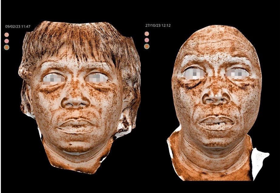
Figure 3 A: Evolution of the patient's skin after one treatment (T1 27/10/23 , after 9 months) compared to baseline (T0 09/02/23) was considered.
Pigmentary component and vascular expression of malar area underwent an initial improvement, but after the summer season a slight worsening with mild post-inflammatory hyperpigmentation.
A second treatment was performed at T1.

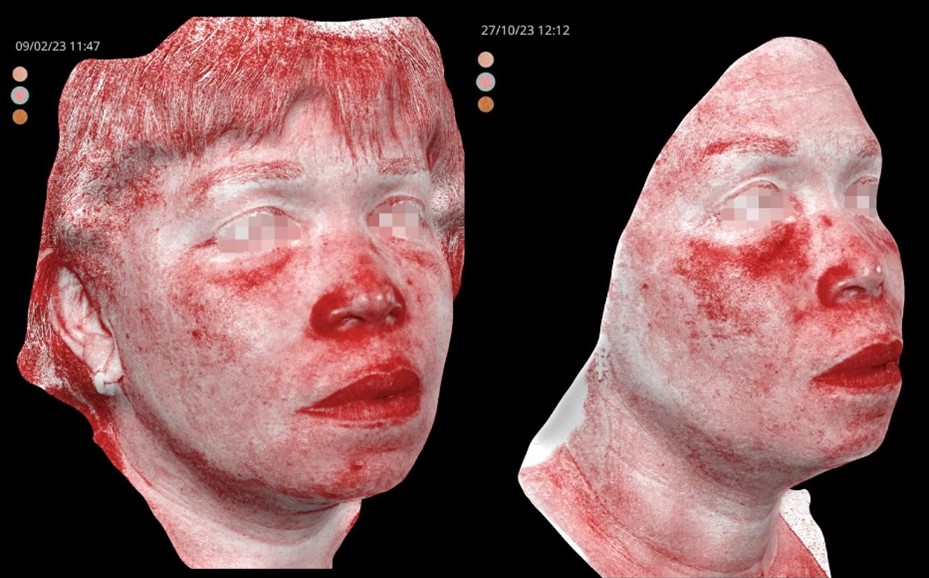
Figure 3B: Evolution of the patient's skin after one treatment (T1 27/10/23 , after 9 months) compared to baseline (T0 09/02/23) was considered.
Pigmentary component and vascular expression of malar area underwent an initial improvement, but after the summer season a slight worsening with mild post-inflammatory hyperpigmentation.
A second treatment was performed at T1.

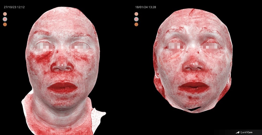
Figure 4: The improvement of the patient's skin after the last treatment (T2 18/01/24, after 11 months) compared to baseline (T0 09/02/23) was considered.
The post-inflammatory hyperpigmentation after T1, was effectively treated after a second treatment.

Figure 5: T2 final result after two treatments.

Figure 6: T0 baseline.

Figure 7: T0-T2.

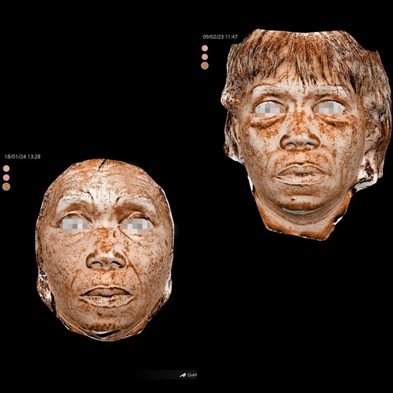
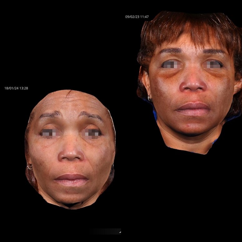
Figure 8: The improvement of the patient's skin after the last treatment (T2 18/01/24, after 11 months) compared to baseline (T0 09/02/23) was considered.

Figure 9: T1-T2.

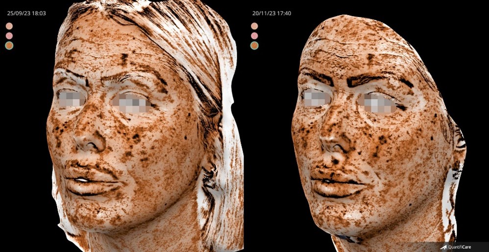
Figure 10A: T0-T1.

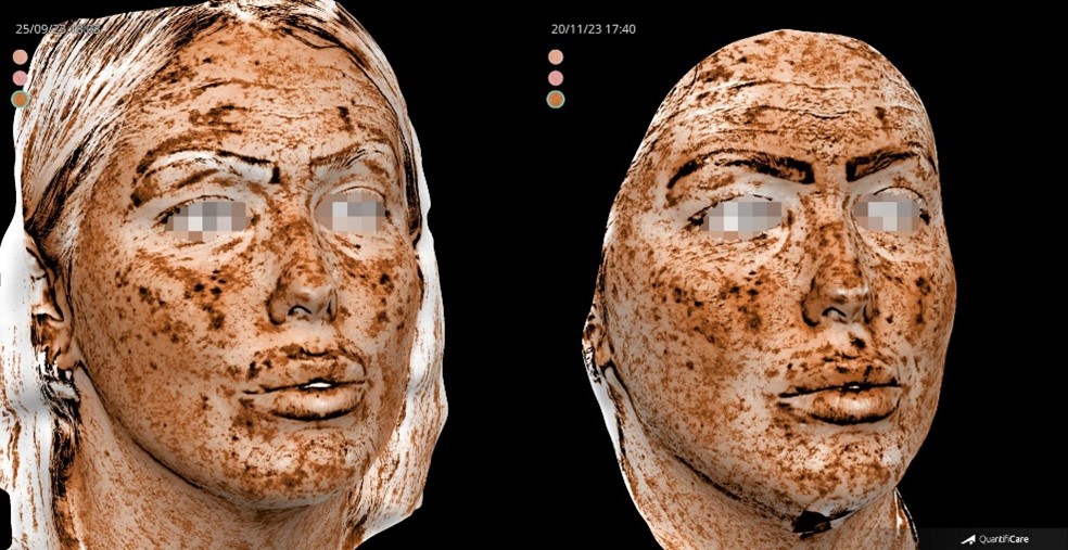
Figure 10B: T0-T1.

Figure 11A: T0-T1.

Figure 11B: T0-T1.

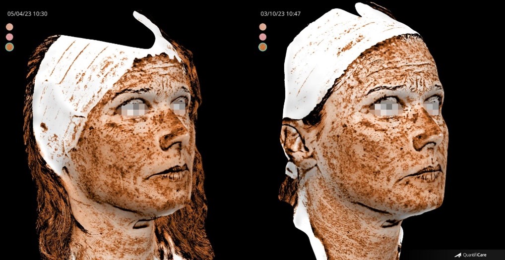
Figure 11C: T0-T1.

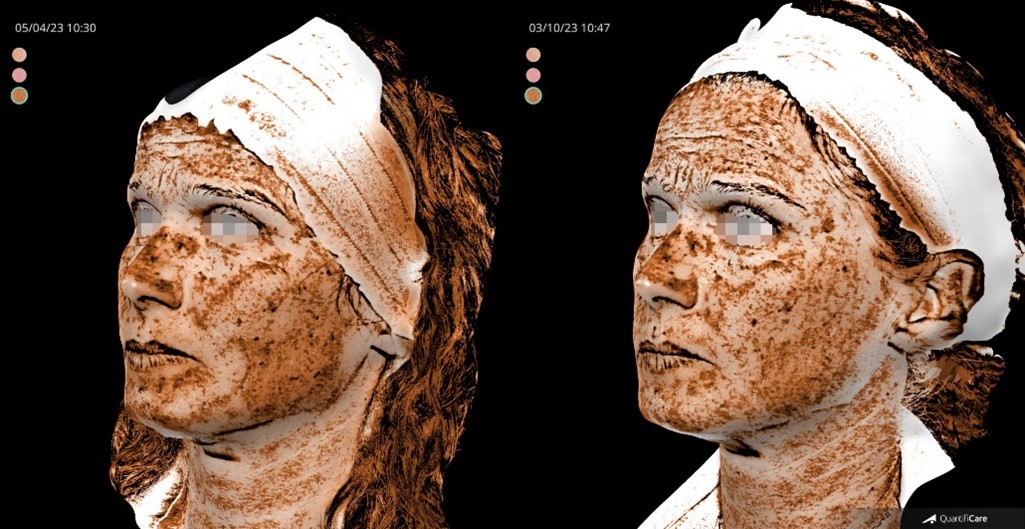
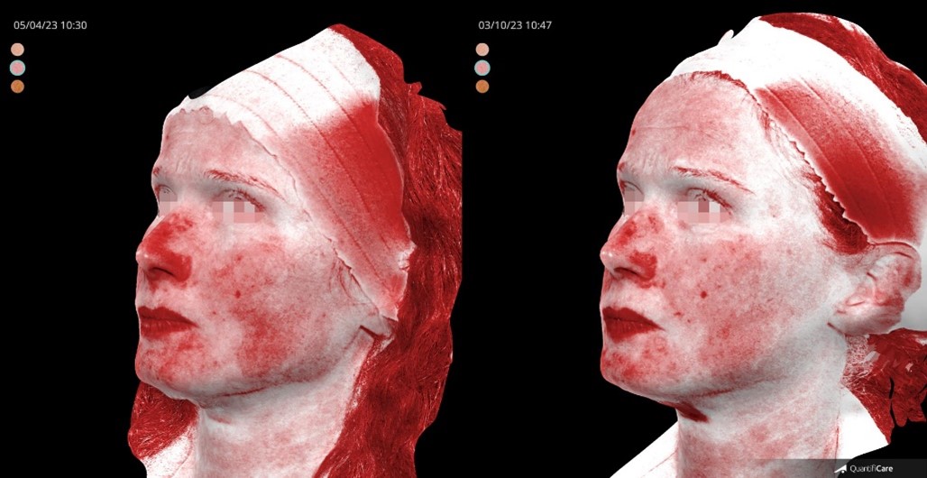
Figure 11D: T0-T1.

Figure 11E: T0-T1.
Results
For all patients, it was possible to evaluate skin improvement before and after the last treatment with RedTouchPro. In general, at T1 a consistent improvement in the pigmentary and vascular components was visible.
All patients tolerated treatment well. Most patients reported pain associated with the procedure as mild.
Common results
Patient's skin improvement after a single session (T1 03/10/23). A remarkable reduction of the entire diffused pigmentation.
Furthermore, considering the outcomes achieved. All patients experienced enhancements in skin texture, thickness, brightness, and overall skin health.
Conclusion
Novel 675 nm laser may be useful for treating benign pigmented lesions because it has a high affinity for melanin and minimal interaction with the vascular component.
The purpose of this case series is to support the scientific literature on the application of a 675-nm laser device for the treatment of vascular and pigmentary symptoms associated with facial melasma in both men and women. The author believes that the laser can be very effective for benign pigmented lesions, lowering the risk of side effects and simplifying post-treatment management because of its strong affinity for collagen and melanin as well as the typical anatomical capillary structure.
Conflicts of interest: The author has no conflicts of interest.
Grant Information: The author received no specific funding for this work.
References
- Ogbechie-Godec OA, Elbuluk N. Melasma: an up-to-date comprehensive review. Dermatol Ther (Heidelb), 2017; 7(3): 305-318. doi:10.1007/S13555-017-0194-1
- Pichardo R, Vallejos Q, Feldman SR, et al. The prevalence of melasma and its association with quality of life in adult male Latino migrant workers. Int J Dermatol, 2009; 48(1): 22-26. doi:10.1111/J.1365-4632.2009.03778.X
- Sarkar R, Gokhale N, Godse K, et al. Medical management of melasma: a review with consensus recommendations by Indian Pigmentary Expert Group. Indian J Dermatol, 2017; 62(6): 450-469. doi:10.4103/IJD.IJD_489_17
- Liu W, Chen Q, Xia Y. New mechanistic insights of melasma. Clin Cosmet Investig Dermatol, 2023; 16: 429-442. doi:10.2147/CCID.S396272
- Espósito MCC, Espósito ACC, Jorge MFS, D'Elia MPB, Miot HA. Depression, anxiety, and self-esteem in women with facial melasma: an Internet-based survey in Brazil. Int J Dermatol, 2021; 60(9): e346-e347. doi:10.1111/IJD.15490
- Nijhawan RI, Sharon JE, Woolery-Lloyd H. Skin of color education in dermatology residency programs: Does residency training reflect the changing demographics of the United States?. Journal of the American Academy of Dermatology, 2008; 59(4): 615-618. doi:10.1016/j.jaad.2008.06.024
- Handel A, Miot L, Miot H. Melasma: A clinical and epidemiological review. An Bras Dermatol, 2014; 89(5): 771-782. doi:10.1590/abd1806-4841.20143063
- Neagu N, Conforti C, Agozzino M, Marangi GF, Morariu SH, Pellacani G, et al. Melasma treatment: a systematic review. J Dermatolog Treat, 2022; 33(4): 1816-1837. doi: 10.1080/09546634.2021.1914313.
- Bailey AJM, Li HO, Tan MG, Cheng W, Dover JS. Microneedling as an adjuvant to topical therapies for melasma: A systematic review and meta-analysis. J Am Acad Dermatol, 2022; 86(4): 797-810. doi: 10.1016/j.jaad.2021.03.116.
- Trivedi MK, Yang FC, Cho BK. A review of laser and light therapy in melasma. Int J Womens Dermatol, 2017; 3(1): 11-20. doi:10.1016/J.IJWD.2017.01.004
- Cannarozzo G, Silvestri M, Tamburi F, et al. A new 675-nm laser device in the treatment of acne scars: an observational study. Lasers Med Sci, 2021; 36(1): 227-231. doi:10.1007/S10103-020-03063-6 doi:10.1089/PHOTOB.2020.4908
- Taroni P, Paganoni AM, Ieva F, et al. Non-invasive optical estimate of tissue composition to differentiate malignant from benign breast lesions: a pilot study. Sci Rep, 2017; 7: 40683. doi:10.1038/SREP40683
- Cannarozzo G, Bennardo L, Zingoni T, Pieri L, Del Duca E, Nisticò SP. Histological skin changes after treatment with 675-nm laser. Photobiomodul Photomed Laser Surg, 2021; 39(9): 617-621. doi:10.1089/PHOTOB.2020.4927
- Bonan P, Verdelli A, Pieri L, Fusco I. Could 675-nm laser treatment be effective for facial melasma even in darker phototype? Photobiomodul Photomed Laser Surg, 2021; 39(10): 634-636. doi:10.1089/PHOTOB.2021.0076
- Nisticò SP, Tolone M, Zingoni T, et al. A new 675-nm laser device in the treatment of melasma: results of a prospective observational study. Photobiomodul Photomed Laser Surg, 2020; 38(9): 560-564. doi:10.1089/PHOTOB.2020.4850

