Colonic Lipomas: An Unusual Cause of Abdominal Pain
Jidal M*, El Bqaq I, Saouab R and El Fenni J
Radiology department, Mohammed V military hospital of Rabat, Morocco
Received Date: 12/12/2023; Published Date: 09/05/2024
*Corresponding author: Jidal M, Radiology department, Mohammed V military hospital of Rabat, Morocco
Lipomas of the gastrointestinal tract have been previously described as quite uncommon, slow-growing, non-epithelial benign adipose tumors that can occur anywhere along the gut. Although they are solitary more often than not, cases of multiple lipomas can be seen in 6-25% [1].
Colonic lipomas were described for the first time by Bauer in 1757. After hyperplastic and adenomatous polyps, they are considered to be the third most common benign colonic tumor. Peak occurs between the fifth and seventh decade, with a female preponderance, and incidence varies between 0.2 and 4.4%. At least, 70% to 90% occur in the right colon [1].
Most of these tumors are submucosal, with the remaining originating from the subserosal or intramucosal area. And since they are usually small, they are most often than not asymptomatic. Symptoms start when the size exceeds 2cm, and include abdominal pain, diarrhea, constipation, blood loss, and intusseption [2].
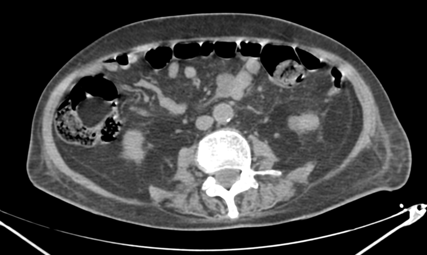
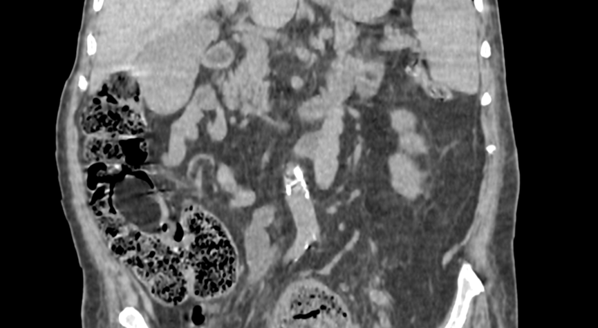
CT is considered to be a good tool to assess the fat content of lipomas and therefore identify a mass as a lipoma. The findings are a homogenous, sharply marginated intraluminal mass with usually an ovoid shape and a characteristic fat density between -40 and -120 Hounsfield units [2,3] (Figure 1, 2). Some studies revealed the possibility of the mass containing linear strands of soft tissue at its base, corresponding on pathologic examination to fibrovascular septas enlarged by inflammation associated with an ulcer [2,3]. The thickness of the septas can help distinguish lipomas from liposarcomas, which are difficult to differentiate [3].
Colonoscopy allows direct visualization of the submucosal lipoma and can help differentiate colonic lipomas from cancer or other tumors. It may also show ulcerated or necrotic mucosa covering the lesion [4].
Lipoma treatment involves two options: endoscopic resection or surgical resection.
Symptomatic and large lipomas should be treated. If there is uncertainty in diagnosis, small and asymptomatic lipomas can be removed. If there is no doubt in the diagnosis, it is reasonable to withhold treatment for asymptomatic lipomas [4].


Figure 1 and 2: Axial and coronal views showing an ovoid well demarcated intraluminal mass in the right colon, with fat density (-111 UH), measuring 32x27x47mm (TxAPxH), in a 57 y.o patients who presented with abdominal pain.
References
- Ozen O, Guler Y, Yuksel Y. Giant colonic lipoma causing intussusception: CT scan and clinical findings. Pan Afr Med J, 2019; 32: 27. doi: 10.11604/pamj.2019.32.27.18040.
- Taylor AJ, Stewart ET, Dodds WJ. Gastrointestinal lipomas: a radiologic and pathologic review. American Journal of Roentgenology, 1990; 155(6): 1205–1210. doi:10.2214/ajr.155.6.2122666
- Thompson WM. Imaging and Findings of Lipomas of the Gastrointestinal Tract. American Journal of Roentgenology, 2005; 184(4): 1163–1171. doi:10.2214/ajr.184.4.0184116
- Goasguen N, Cattan P, Godiris-Petit G, Munoz-Bongrand N, Allez M, Lemann M, et al. Lipome colique: cas clinique et revue de la littérature. Gastroentérologie Clinique et Biologique, 2008; 32(5): 521–524. doi:10.1016/j.gcb.2007.11.007

