Squamous Cell Carcinoma: Unusual Dermoscopic Appearance
Mejjati Kaoutar1,*, Douhi Zakia1, Soughi Meryem1, Elloudi Sara1, Baybay Hanane1, Layla Tahiri Elousrouti2 and Mernissi Fatima Zahra1
1Department of Dermatology, University Hospital Hassan II, Morocco
2Pathology Department, University Hospital Hassan II, Morocco
Received Date: 03/11/2023; Published Date: 09/04/2024
*Corresponding author: Mejjati Kaoutar, Department of Dermatology, University Hospital Hassan II, Fez, Morocco
Case Presentation
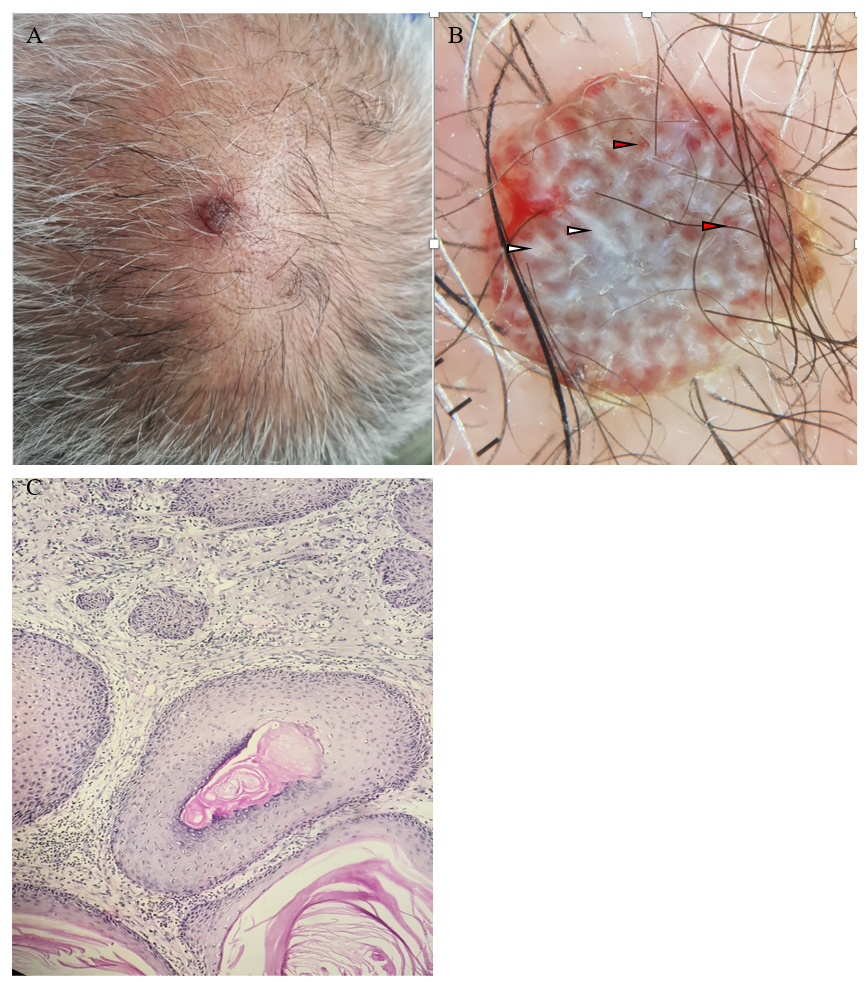
A 72-year-old patient, consulted for a lesion on the scalp that had been evolving for 2 months. Dermatological examination revealed a purplish-grey rounded nodule, measuring approximately 2 cm, with a smooth surface bleeding on contact (Figure 1A). Dermoscopy revealed shine white streaks and pseudolacunae containing irregular vessels (Figure 1B).
The patient underwent an excision of the nodule revealing a squamous proliferation made up of polygonal cells with atypical nuclei, and cystic areas containing keratinous material confirming the clinical diagnosis of a mature, well differentiated, keratinizing and infiltrating squamous cell carcinoma (Figure 1C).
Considering clinical and histopathologic findings, diagnosis of carcinoma cuniculatum (CC) was made.

Figure 1: (A) rounded reddish-purple nodule on the head (B) Dermoscopy of the lesion showing pseudolacunae (red arrow ) and chrysalis ( white arrow ) (C) Dermal infiltration by squamous proliferation arranged in large, sometimes encysted clusters , filled with concentric keratin lamellae (HES x200).
Teaching point:
CC is a variant of squamous cell carcinoma, most often found on the plantar surface, but also on the head. Histologically, it is characterized by the presence of interconnected keratin-filled crypts of a well-differentiated squamous cell carcinoma with a predominantly endophytic growth pattern (1). Previous studies have described the dermoscopic appearance of squamous cell carcinoma: scales, blood spots, white circles, white structureless areas, hairpin vessels, linear-irregular vessels, perivascular white halos, and ulceration. Chrysalis have rarely been reported in squamous cell carcinoma (2). Their abundance in our case could be correlated histologically to the keratin contained in the cysts, which are characteristic of CC. Through our observation, we would like to draw attention to this dermoscopic aspect of the CC.
References
- Fania L, Didona D, Di Pietro FR, Verkhovskaia S, Morese R, Paolino G, et al. Cutaneous Squamous Cell Carcinoma: From Pathophysiology to Novel Therapeutic Approaches. Biomedicines, 2021; 9(2): 171. doi: 10.3390/biomedicines9020171. PMID: 33572373; PMCID: PMC7916193
- Balagula Y, Braun RP, Rabinovitz HS, Dusza SW, Scope A, Liebman TN, et al. The significance of crystalline/chrysalis structures in the diagnosis of melanocytic and nonmelanocytic lesions. J Am Acad Dermatol, 2012; 67(2): 194.e1-8. doi: 10.1016/j.jaad.2011.04.039. Epub 2011 Oct 24. PMID: 22030020.

