Coil Embolization for Septal Ablation in a Young Adolescent with Hypertrophic Obstructive Cardiomyopathy
You-Jyun Jhou1, Li-Han Chen1,2, Ching-Tang Chang1,2, Tzu-chieh Lin1,2, Ho-Ming Su1,2,3, Tsung-Hsien Lin1,2,3, Nai-Yu Chi1,2,* and Po-Chao Hsu1,2,3,*
1Department of Internal Medicine, Kaohsiung Medical University Hospital, Kaohsiung Medical University, Kaohsiung, Taiwan
2Division of Cardiology, Department of Internal Medicine, Kaohsiung Medical University Hospital, Kaohsiung Medical University, Kaohsiung, Taiwan
3Department of Internal Medicine, Faculty of Medicine, School of Medicine, Kaohsiung Medical University, Kaohsiung, Taiwan
Received Date: 31/08/2023; Published Date: 19/01/2024
*Corresponding author: Nai-Yu Chi & Po-Chao Hsu, Division of Cardiology, Department of Internal Medicine; Kaohsiung Medical University Hospital, 100 Tzyou 1st Road, Kaohsiung. 80708, Taiwan, ROC
Abstract
Hypertrophic Obstructive Cardiomyopathy (HOCM) is a common genetic cardiac disorder characterized by left ventricular hypertrophy and outflow tract obstruction. Medical therapy is often the first-line treatment for HOCM. However, some patients remain symptomatic despite maximal medical therapy, and alternative interventions such as surgical myectomy and alcohol septal ablation may be needed. However, these procedures are associated with some morbidities. Coil embolization of a septal branch of the left anterior descending artery has emerged as a less invasive alternative that may provide similar benefits with a lower risk of complications. Herein, we presented an 18 years-old adolescent with HOCM successfully treated by coil embolization for septal reduction treatment. To our knowledge, our case should be the youngest case of HOCM treated by coil embolization in the current literature.
Keywords: Hypertrophic obstructive cardiomyopathy; Coil embolization; Left ventricular outflow tract
Introduction
Hypertrophic Cardiomyopathy (HCM) is a genetically determined heart muscle disease, which may develop heart problem such as LVOT obstruction, diastolic dysfunction, myocardial ischemia, or mitral valve regurgitation [1]. These structural and functional abnormalities cause symptoms of exertional dyspnea, chest pain, fatigue, palpitations, syncope, or even sudden cardiac death [2]. Left Ventricular Outflow Tract (LVOT) obstruction is a strong, independent predictor of heart failure (HF) symptoms. Treatment strategies attempt to increase LV chamber size or decrease cardiac inotropy, thereby diminishing Systolic Anterior Motion (SAM)-septal contact, resulting in a reduced or abolished LVOT gradient. Pharmacologic therapy is the first-line treatment strategy in patients with HCM and symptomatic LVOT obstruction [3]. For patients with symptoms/signs of heart failure (New York Heart Association class III/IV) persist despite maximal medical therapy, patients have recurrent syncope judged to be related to hemodynamic compromise from LVOT obstruction, or an LVOT gradient ≥50 mmHg is present at rest or with provocation, invasive septal reduction therapies may be indicated [4]. In recent years, coil embolization is a new choice different from surgical myectomy or alcohol septal ablation [5-7]. However, there are limited cases discussing about coil embolization performed in a young adolescent. Herein, we presented a 18 years-old cases of hypertrophic obstructive cardiomyopathy (HOCM) successfully treated by coil embolization for septal reduction treatment.
Case Presentation
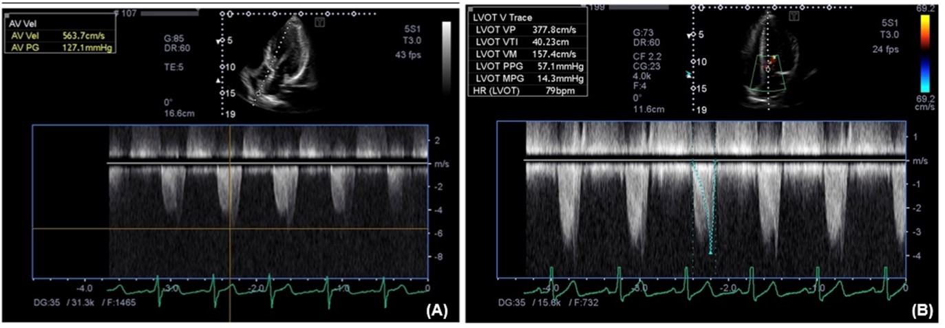
An 18 years-old young adolescent, who denied of systemic disease before, was presented with decreasing effort and deterioration of dyspnea on exertion in recently half year. 12-lead electrocardiogram revealed left ventricular hypertrophy with high voltage in multiple leads (Figure 1). Echocardiogram showed HCM with LVOT obstruction (LVOT pressure gradient 127 mmHg) (Figure 2A). In addition, SAM (systolic anterior motion) was also found. Due to symptomatic HCM with highly LVOT pressure gradient and SAM, invasive septal reduction therapy was indicated. After discussing with patient, he wanted to received coil embolization for septal reduction therapy. Cardiac computed tomographic angiography was performed and no evidence of congenital anomaly and obstruction of coronary artery was noted. In addition, the largest septal branch of left anterior descending artery was the second one. During catheterization, coil embolization for septal branch of left anterior descending artery was performed and finally 7 coils were used (Figure 3).
After the procedure, patient was transferred to intensive care unit for further care. Cardiac enzymes were peaked as: Troponin I: 1.16 ng/ml, CKMB: 11.4 ng/ml, CPK: 281 IU/L. Pressure gradient of LVOT measured before catheterization was 127 mmHg. After embolization, pressure gradient gradually decreased to 57mmHg after several days later (Figure 2B). In addition, non-sustained ventricular tachycardia was noted on the day 1 after embolization, and returned to normal sinus rhythm after verapamil use. This patient was discharged uneventfully.

Figure 1: 12-lead electrocardiogram revealed left ventricular hypertrophy with high voltage in multiple leads.

Figure 2: Pressure gradient over left ventricular outflow tract (LVOT).
Figure 2A: Pressure gradient of LVOT before coil embolization (127mmHg).
Figure 2B: Pressure gradient of LVOT post coil embolization (57mmHg).

Figure 3: Step by step of Coil embolization during catheterization (From A to F).
Discussion
Coil embolization of the septal artery has emerged as a novel treatment option for selected patients with symptomatic HOCM. The technique involves the deployment of embolic coils in the septal artery, leading to localized ischemia and subsequent reduction in left ventricular outflow tract obstruction [5-7]. The procedure can be performed under conscious sedation or general anesthesia, and is associated with low morbidity and mortality rates. However, careful patient selection and experienced operators are crucial for optimal outcomes. Coil embolization can provide effective symptomatic relief, with significant improvements in quality of life, exercise tolerance, and left ventricular function. Furthermore, coli embolization needs time for ischemic efforts of septal wall, reduction of LVOT pressure gradient could not be detected immediately after coli embolization. In our case, we found the decreased pressure gradient of LVOT (57mmHg) around several days later after coil embolization. We also arranged strain echocardiogram for left ventricular segmental wall motion and it showed impaired systolic function of septal wall near LVOT, which represented successful embolization by coil embolization.
Conclusion
Coil embolization of the septal artery is a less invasive alternative to surgical myectomy for selected patients with symptomatic HOCM. The procedure is safe and effective, providing significant improvements in symptoms, quality of life, and left ventricular function. Careful patient selection and experienced operators are crucial for optimal outcomes. To our knowledge, our case should be the youngest case of HOCM treated by coil embolization in the current literature. This case reminds physicians that coil embolization is also a possible and effective treatment for young adolescent with HOCM.
References
- Maron MS, Olivotto I, Zenovich AG, et al. Hypertrophic cardiomyopathy is predominantly a disease of left ventricular outflow tract obstruction. Circulation, 2006; 114(21): 2232-2239.
- Adabag AS, Casey SA, Kuskowski MA, et al. Spectrum and prognostic significance of arrhythmias on ambulatory Holter electrocardiogram in hypertrophic cardiomyopathy. J Am Coll Cardiol, 2005; 45(5): 697.
- Veselka J, Anavekar NS, Charron P. Hypertrophic obstructive cardiomyopathy. Lancet, 2017; 389(10075): 1253-1267.
- American College of Cardiology Foundation/American Heart Association Task Force on Practice Guidelines; American Association for Thoracic Surgery; American Society of Echocardiography; American Society of Nuclear Cardiology; Heart Failure Society of America; Heart Rhythm Society; Society for Cardiovascular Angiography and Interventions, et al. 2011 ACCF/AHA guideline for the diagnosis and treatment of hypertrophic cardiomyopathy: executive summary: a report of the American College of Cardiology Foundation/American Heart Association Task Force on Practice Guidelines. J Thorac Cardiovasc Surg, 2011; 142(6): 1303-1338.
- Guerrero I, Dhoble A, Fasulo M, et al. Safety and efficacy of coil embolization of the septal perforator for septal ablation in patients with hypertrophic obstructive cardiomyopathy. Catheter Cardiovasc Interv, 2016; 88(6): 971-977.
- Song JH, Park SH, Song HJ, et al. Coil Embolization of Septal Branches in Hypertrophic Obstructive Cardiomyopathy. Korean Circulation J, 2004; 34: 706-710.
- Durand E, Mousseaux E, Coste P, et al. Non-surgical septal myocardial reduction by coil embolization for hypertrophic obstructive cardiomyopathy: early and 6 months follow-up. Eur Heart J, 2008; 29(3): 348-355.

