General MRI Features of Focal Cortical Dysplasia
Kaouthar Sfar*, Nada Adjou, Kaoutar Maslouhi, Fatima Chait, Nazik Allali, Siham El Haddad, Latifa Chat
Mother and Child Imaging Department, HER – Ibn Sina Hospital, Mohammed V University in Rabat, Rabat, Morocco
Received Date: 05/06/2023; Published Date: 13/10/2023
*Corresponding author: Kaouthar Sfar, Mother and Child Imaging Department, HER – Ibn Sina Hospital, Mohammed V University in Rabat, Rabat, Morocco
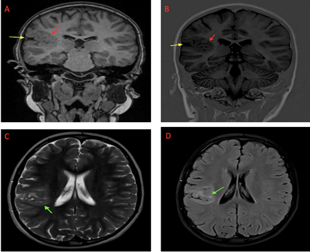
Focal cortical dysplasia is one of the most common causes of drug-resistant partial epilepsy in the pediatric population [3]. This term has been used to refer to a heterogenous group of focal disorders of the cortical mantle development [3]. Several classifications, based on histopathological, neuroimaging, and genetic features, have been proposed to distinguish between the different subtypes. Nevertheless, there is an overlap between the MRI brain findings of the different types. General imaging features (Figure) include [1,2]:
- Focal cortical thickening and dysmorphia.
- Blurring of the gray matter-white matter junction.
- Increased T2 and FLAIR signal intensity of gray matter and subcortical white matter with or without transmatle sign (40%).
- In T1- weighted sequences, the signal intensity of the subcortical white matter decreases.
- There is no contrast enhancement.
- Abnormal sulcal.
- Focal brain hypoplasia: loss of lobar or sublobar volume with enlargement of subarachnoid space.

Figure: MRI images of focal cortical dysplasia in a 10 years old boy with a status epilepticus. Coronal T1 (A) and turbo spin-echo inversion-recovery T1-weighted (B) sequences exhibit a focal cortical dysplasic thickening with blurring between GM/WM junction (à), enlarged subarachnoid space and abnormal sulcal (à). Axial T2 and FLAIR show hyperintensity of gray matter and subcortical white matter (à).
References
- Colombo N, Tassi L, Galli C, et al. Focal cortical dysplasias: MR imaging, histopathologic, and clinical correlations in surgically treated patients with epilepsy. AJNR, 2003; 24: 724–733.
- Celi Santos Andrade, Claudia da Costa Leite. Malformations of cortical development: Current concepts and advanced neuroimaging review. Arquivos de Neuro-psiquiatria, 2011; 69(1): 130-138.
- Kabat J, Krol P.Focal cortical dysplasia – review.Pol J Radiol, 2012; 77(2): 35–43.

