A Magic Chest X-Ray: The Chinese Rings
Otman Bouzouba*, Abdelouahhab Bazsalt and Abla Khellaf
Resident, Cardiogeriatrics Department, René-Muret Hospital, APHP, Sevran, France
Received Date: 30/12/2022; Published Date: 23/01/2023
*Corresponding author: Otman Bouzouba, Resident, Cardiogeriatrics Department, René-Muret Hospital, APHP, Sevran, France
Clinical Case
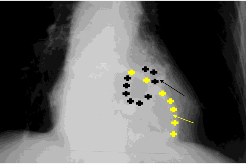
Mrs. B, 86 years old, was hospitalized in the service for assessment of falls. The history of the falls was in favor of balance disorders, which were confirmed by the existence of a cerebellar syndrome at admission. Mrs. B. had no particular history and was not taking any medication. Cardiovascular examination was normal at admission. A routine chest X-ray showed the following appearance (Figure 1).

Figure 1: Frontal chest x-ray: Image evoking a Chinese ring formed by the intersection of the calcified mitral annulus (black arrow, drawn by black plus sign) and an annulus formed by a large hiatal hernia (yellow arrow, drawn by yellow plus sign).

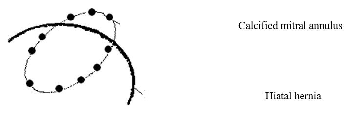
Figure 2: Figures 1 et 2: Entanglement of two elliptical intersecting perpendicularly by the major axis.

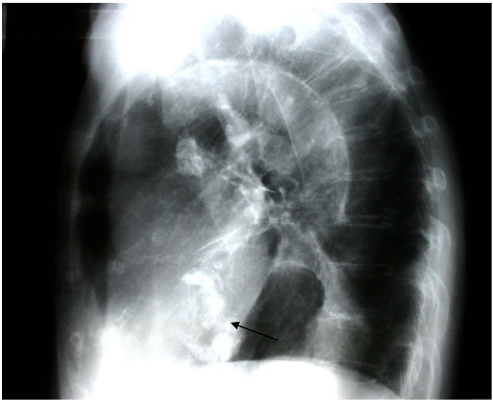
Figure 3: Left profile chest x-ray: Elliptical ring which major axis is oriented upwards and forwards (Arrow).

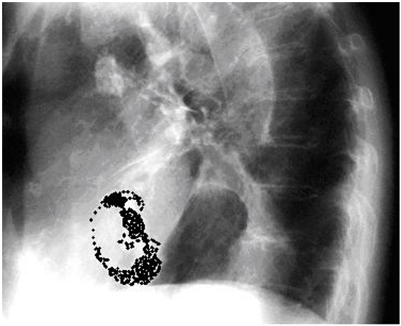
Figure 4: Left profile chest x-ray: visualization of the calcified mitral annulus by redrawing it.
Discussion
The discovery of a calcified mitral annulus in an elderly subject is mainly the witness of a general atheromatous disease, as evidenced by the publications of Ix and Sgorbini [1,2] and its possible complications [3]. However, an observation concerning a young patient with Marfan's disease also reported the existence of massive calcification of the mitral annulus [4].
By retrospectively analyzing 24,380 echocardiograms, Mohaved et al [5] showed a prevalence of 6.1% of mitral annulus calcifications. These calcifications were more frequently found in cases of mitral regurgitation, tricuspid regurgitation, left ventricular hypertrophy and aortic stenosis, compared to patients without these valvular and/or cardiac structural anomalies.
The shape of the calcified annulus and its anatomical relationships confirm that it is a calcified mitral annulus. The second larger diameter annulus visible only from the front represents the gastric mucosa of a large hiatal hernia.
It should be noted that the biology of this patient was normal, in particular the renal function and the calcemia even corrected.
Funding: The authors declare that no funding was received for this article;
Declaration of Competing Interest: None of the authors have any conflicts of interest to disclosure
Authors Contribution:
Otman Bouzouba: acquisition of data, drafting the article and literature revision, guarantor
Abdelouhhab Bazzout: acquisition of data and literature revision
Abla Khellaf: critical revision
References
- Ix JH, Chertow GM, Shlipak MG, et al. Association of fetuin-A with mitral annular calcification and aortic stenosis among persons with coronary heart disease: data from the Heart and Soul Study. Circulation, 2007; 115(19): 2533-2539.
- Sgorbini L, Scuteri A, Leggio, et al. Carotid intima-media thickness, carotid distensibility and mitral, aortic valve calcification: a useful diagnostic parameter of systemic atherosclerosis disease. J Cardiovasc Med, 2007; 8(5): 342-347.
- Poh KK, Wood MJ, Cury RC. Prominent posterior mitral annular calcification causing embolic stroke and mimicking left atrial fibroma. Eur Heart J, 2007.
- Correia J, Rodrigues D, Da Silva AM, et al. Massive calcification of the mitral valve annulus in an adolescent with Marfan syndrome. A case reports. Rev Port Cardiol, 2006; 25(10): 921-926.
- Mohaved MR, Saito Y, Ahmadi-Kashani M, Ebrahimi R. Mitral annulus calcification is associated with valvular and cardiac structural abnormalities. Cardiovasc Ultrasoud, 2007; 14(5): 14.

